

Introduction
Materials and Methods
Chemicals
Preparation of a 70% ethanol extract of A. tenuissima root (ATE)
HPLC Analysis
Determination of total phenolic and total flavonoid content in ATE
Effect of ATE on DPPH radical scavenging activity
Effect of ATE on ABTS radical scavenging activity
Determination of the reducing power of ATE
Cell culture
Cell viability
Effect of ATE on the production of melanin in B16F10 cells
Tyrosinase activity assay
Effect of ATE on the in vitro inhibitory activity of tyrosinase
Effect of ATE on the in vitro activity of α-glucosidase activity
Statistical Analysis
Results
Determination of content of decursin and Z-ligustilide in ATE
The content of total phenolic and total flavonoid
Effect of ATE on the cell viability
Antioxidant capacity of ATE
Inhibitory effect of ATE on the melanin production in B16F10 cells
Effect of ATE on the in vitro tyrosinase and α-glucosidase activities
Discussion
Introduction
Angelica tenuissima Nakai (= Ligusticum tenuissimum Kitagawa) belongs to the family Umbelliferae and is distributed in certain areas in China and rocky slopes in the Korean peninsula (Ka et al., 2005). The plant grows to a height of 30-80 ㎝, has no hair throughout thereof and emits fragrance. The plant bears oval fruits, and the root thereof is used for medicinal purposes. The root gathered in the season of fall has been used as traditional medicines treating headache and remedies for women with gynecological diseases and anemia in Asia (Kim et al., 1997; Han et al., 1998). Recently, interferon-mediated anti-viral, anti- osteoporosis, and anti-inflammatory activities of A. tenuissima have been identified (Lee et al., 2010; Ahn et al., 2015; Weeratunga et al., 2016). Various types of compounds, including decursin, Z-ligustilide, ferulic acid, and 3-butylidene-4,5- dihydrophthalide have been identified in A. tenuissima (Ka et al., 2005). Although studies on the separation and identification of the components of A. tenuissima have been made, there are few relevant reports on its biochemical activities currently.
The melanin pigment is a polymer produced inside the melanosomes under the influence of tyrosinase, which converts L-tyrosine to dopaquinone during melanogenesis (Smit et al., 2009; Skoczynska et al., 2017). Numerous factors, such as ultraviolet light (UV), inflammation, and rubbing of the skin as well as abnormal α-melanocyte stimulating hormone (α-MSH) release, are involved in the progression of pigmentary disorders (Bastonini et al., 2016). Excessive production of cutaneous melanin may cause considerable problems of esthetic nature, especially in hyperpigmentary conditions, like melasma, age spots, postinflammatory hyperpigmentation, freckles or lentigines (Smit et al., 2009). The basic molecular mechanism of skin whitening is aimed at reduction of the melanin production (Smit et al., 2009). Development of preparations for bleaching hyperpigmented lesions or to safely achieve overall whitening is one of the challenges for cosmetic industry, especially in East Asia.
The skin is continuously exposed to reactive oxygen species (ROS) endogenous such as inflammation and exogenous sources including UV and environmental pollutants. Oxidative stress plays a crucial role in upregulated synthesis of melanin and thus accelerates skin pigmentation (Masaki, 2010). Moreover, topical application of antioxidants provide potential benefits for skin as reviewed elsewhere (Silva et al., 2017). Skin ageing, such as a wrinkle formation and abnormal pigmentation, is also associated with skin damage caused by endogenous reactive oxygen species (ROS) (Masaki, 2010). Hence, the suppression of oxidative stress in the skin will be favorable in reduction and/or prevention of atypical pigmentation. Therefore, in addition to single agent which aims at inhibiting tyrosinase, the use of complex mixtures that target different mechanism like anti-oxidant and anti- inflammatory effects have been undergoing (Smit et al., 2009; Bin et al., 2016).
Although A. tenuissima has been used widely in treating headache, diarrhea, epilepsy and rheumatic arthralgias (Kim et al., 1997; Han et al., 1998), its anti-melanogenic effect has not been reported in detail. Hence, in the present study, anti- oxidative potential of 70% ethanol extract of A. tenuissima (ATE) was determined by DPPH and ABTS scavenging activities, and total reducing potential power assay which is one of important factors led to excessive melanin production in the skin. Moreover, the anti-melanogenic potential of ATE on B16F10 cells was investigated through its inhibitory activity on the melanin production which is highly associated with tyrosinase and/or α-glucosidase activity.
Materials and Methods
Chemicals
2,2-diphenyl-1-picryl-hydrazyl-hydrate (DPPH), β-phycoerythrin, mushroom tyrosinase, and α-melanocyte stimulating hormone (α-MSH), decursin, Z-ligustilide, dimethylsulfoxide (DMSO), L-3,4-dihydroxyphenylalanine (L-DOPA), and deionized distilled water were purchased from Sigma (St Louis, MO, USA). Dulbecco's modified eagle medium (DMEM) and fetal bovine serum (FBS) were obtained from Gibco (NY, USA). Unless indicated otherwise, all other chemicals were obtained from Sigma.
Preparation of a 70% ethanol extract of A. tenuissima root (ATE)
A. tenuissima Nakai roots were purchased from Jaechun oriental medicine market and identified by Dr. S.C. Kang of Kyung Hee University. Voucher specimens have been deposited at the herbarium of the School of Medicine, Kyung Hee University. Dried roots of A. tenuissima were chopped into small pieces and extracted in 70% ethanol at 80℃ for 4 h. The extract was subsequently filtered to remove any particulates, and was concentrated under vacuum at 50℃ to be 53 brix of solution. Then, the extract was lyophilized to obtain a powder and stored at -20℃ for further experiments. The yield of ATE was 22% (weight/weight) from a dried root of A. tenuissima Nakai.
HPLC Analysis
HPLC analysis was performed using an Waters 600 HPLC system with a PDA detector and a reversed-phase analytical column (Capcell Pak C18 (2), 3 ㎛, 4.6 × 150 ㎜) at a flow rate 0.8 ㎖/min. HPLC grade acetonitrile was purchased from Samchon Chemical, Korea, and used without further purification. All solvents had formic acid (FA) as acid buffer (0.1% v/v). Each lyophilized sample was dissolved in ethanol fully with sonication for 30 min. Injection volume for the analysis was 15 ㎕ of the 1 ㎎/㎖ concentration sample. For the decursin and Z-ligustilide analysis, methanol and water (1:9, v/v), acetonitrile and water (9:1, v/v) (both with 0.1% FA) were used for the gradient solvent condition. Chromatograph was monitored at UV 310 ㎚. Each of the analysis was repeated for three times.
Determination of total phenolic and total flavonoid content in ATE
Total phenolic content was determined according to the method of Folin-Ciocalteu (Sasidharan et al., 2010). The extract was mixed with Folin-Ciocalteu’s (50 g/100 ㎖), phenol reagent and then, Sodium carbonate (2 g/100 ㎖) was added, and the final volume was made up to 5 ㎖ with deionized water. The mixture was allowed to stand at room temperature for 30 min and the absorbance was measured at 750 ㎚ by UV/VIS spectrophotometer. The concentration of total phenolic content of ATE was calculated from the standard curve of gallic acid and expressed as ㎍ gallic acid equivalent (GAE)/㎎. Total flavonoid content was measured using the aluminum chloride colorimetric method (Chang et al., 2002; Marinova et al., 2005). The extract was mixed with aluminum chloride hexahydrate (10 g/100 ㎖) and then, 95% EtOH and 1M Potassium acetate was added, and the final volume was made up to 5 ㎖ with deionized water. After mixing, the solution was incubated for 40 min at room temperature. The absorbance of the reaction mixtures was measured at 415 ㎚ by UV/VIS spectrophotometer. The concentration of total flavonoid content in the sample was calculated from the standard curve and expressed as ㎍ quercetin equivalent (QE)/㎎ of ATE. All the experiments were carried out, at a least, in triplicate.
Effect of ATE on DPPH radical scavenging activity
Different concentrations of ATE were adjusted at 100 ㎕ with reaction mixture and then reacted with 100 ㎕ of 0.4 mM DPPH (2,2-diphenyl-1-picrylhydrazyl) solution in 99% EtOH. After vigorous shaking, reaction mixtures were allowed to reach a steady state at room temperature for 30 min. Decolorization of DPPH was determined by measuring the absorbance at 540 ㎚ with using a Molecular Devices microplate reader. IC50 concentration required to inhibit DPPH radical formation by 50% was calculated from the graph after plotting inhibition percentage against extract concentration.
Effect of ATE on ABTS radical scavenging activity
The 2,2’-azino-bis (3-ethylbenzthiazoline-6-sulfonic acid (ABTS) antioxidant assay was generated by adding 7.4 mM ABTS to 2.4 mM potassium persulfate and the mixture was left at room temperature for overnight in dark. The ABTS radical cation solution was diluted with distilled water to obtain an absorbance of approximately 1.1 ± 0.02 at 734 ㎚. 400 ㎕ of ATE at various concentrations (0 to 1000 ㎍/㎖) was added to 800 ㎕ of ABTS reagent. Reaction was conducted at room temperature for 10 min in dark, and then absorbance was measured at 734 ㎚ with using UV/VIS spectrophotometer (Shimadzu, Japan). IC50 concentration required to inhibit ABST radical formation by 50% was calculated from the graph after plotting inhibition percentage against extract concentration.
Determination of the reducing power of ATE
Different concentrations of ATE were incubated with potassium ferricyanide (1 g/100 ㎖) and 0.2 M sodium phosphate buffer (pH 6.6) at 50℃ for 20 min the reaction was terminated by adding TCA solution (10 g/100 ㎖), centrifugated at 3000 g for 10 min and the supernatant was mixed with ferric chloride (0.1 g/100 ㎖). Reaction was conducted at room temperature for 10 min. The absorbance measured at 700 ㎚. The extract concentration providing absorbance of 0.5 (EC50) was calculated from the graph of absorbance at 700 ㎚ against extract concentration.
Cell culture
B16F10 (mouse skin melanoma) cells were obtained from Korean cell line bank (Seoul, Korea) and were maintained in DMEM supplemented with 2 mM L-glutamine, 100 IU/㎖ penicillin, 100 ㎍/㎖ streptomycin, and 10% heat-inactivated FBS. Cells were maintained in a humidified atmosphere of 5% CO2 at 37℃.
Cell viability
Cell viability was measured by quantitative colorimetric assay with a 3-(4,5-dimethylthiazol-2-yl)-2,5-diphenyltetrazolium bromide (MTT) as previously reported (Koo et al., 2017). Exponentially growing B16F10 cells were seeded at 1×10⁴ cells/well in 96-well tissue culture plates and treated with different dose of ATE (0-2000 ㎍/㎖) for 24 and 48 h. After incubation of MTT (150 ㎍/㎖) for 4 h, the formazan crystals were dissolved in dimethyl sulfoxide (DMSO), and then measured the optical density at 540 ㎚ using a Molecular Devices microplate reader (Menlo Park, CA). Cytotoxicity was expressed as a percent of untreated control cells.
Effect of ATE on the production of melanin in B16F10 cells
B16F10 cells were treated with α-MSH (100 nM) in the presence or absence of ATE (0 to 1000 ㎍/㎖) for 48 h. The cells were washed with PBS and lysed with lysis buffer containing 50 mM sodium phosphate (pH 6.8) buffer, 1% Triton X-100, and 0.1 mM PMSF. After collecting the cell supernatant by centrifugation, the pellets were dissolved in 1 N NaOH for 1 h at 60℃. The absorbance was measured at 405 ㎚ with a microplate reader.
Tyrosinase activity assay
B16F10 cells were treated with α-MSH (100 nM) in the presence of absence of different concentrations of ATE (0 to 1000 ㎍/㎖) for 48 h. The culture medium was then removed, and the cell was washed with PBS and then lysed with lysis buffer containing 50 mM sodium phosphate (pH 6.8) buffer, 1% Triton X-100 and 0.1 mM PMSF. After collecting cell supernatant with centrifugation, tyrosinase activity was determined by addition of reaction mixture (40 ㎕ of 100 mM sodium phosphate buffer (pH 6.8) and 160 ㎕ of 10 mM L-DOPA) in the presence of cell lysate (40 ㎍) for 1 h. Then absorbance was measured at 490 ㎚ with microplate reader.
Effect of ATE on the in vitro inhibitory activity of tyrosinase
Tyrosinase inhibitory activity was measured by a colorimetric method (Lee et al., 2017). Briefly, 40 ㎕ of mushroom tyrosinase (110 units/㎖) was added to 100 ㎕ of reaction mixture 175 mM sodium phosphate buffer (pH 6.8) and added 10 mM L-DOPA 40 ㎕ in the presence or absence of sample. Ascorbic acid (50 ㎍/㎖) was used as a positive control. The reaction was conducted at 25℃ for 5 min and the absorbance was measured at 490 ㎚. Tyrosinase inhibitory activity (%) was calculated using the following equation: [1-(Sample with enzyme - Sample without enzyme)/(Blank with enzyme - Sample without enzyme)] × 100.
Effect of ATE on the in vitro activity of α-glucosidase activity
The inhibition of α-glucosidase activity was determined using the modified published method (Kim et al., 2000). Briefly, α- glucosidase (Saccharomyces cerevisiae, Sigma-Aldrich, USA) was dissolved in 100 mM of Sodium phosphate buffer (pH 6.8) containing 200 ㎎ of bovine serum albumin (Merck, German). The reaction mixture consisting 10 ㎕ of sample was premixed with 90 ㎕ α-glucosidase (1 unit/㎖). After preincubating at 37℃ for 15 min, 100 ㎕ of 1 mM p-nitrophenyl α-D-glucopyranoside (Sigma-Aldrich, Switzerland) and incubated at 37℃ for 5 min. α-Glucosidase activity was determined spectrophotometrically at 405 ㎚ on spectrophotometer. Quercetin (10 ㎍/㎖) was used as positive control of α-glucosidase inhibitor.
Statistical Analysis
Each experiment was repeated three or four times, and the results of a representative experiment are shown. The results are expressed as means ± SEM and were analyzed using one- Way ANOVA followed by Turkey’s method (Systat Software Inc., San Jose, CA, USA). A statistical probability of p<0.05 was considered significant.
Results
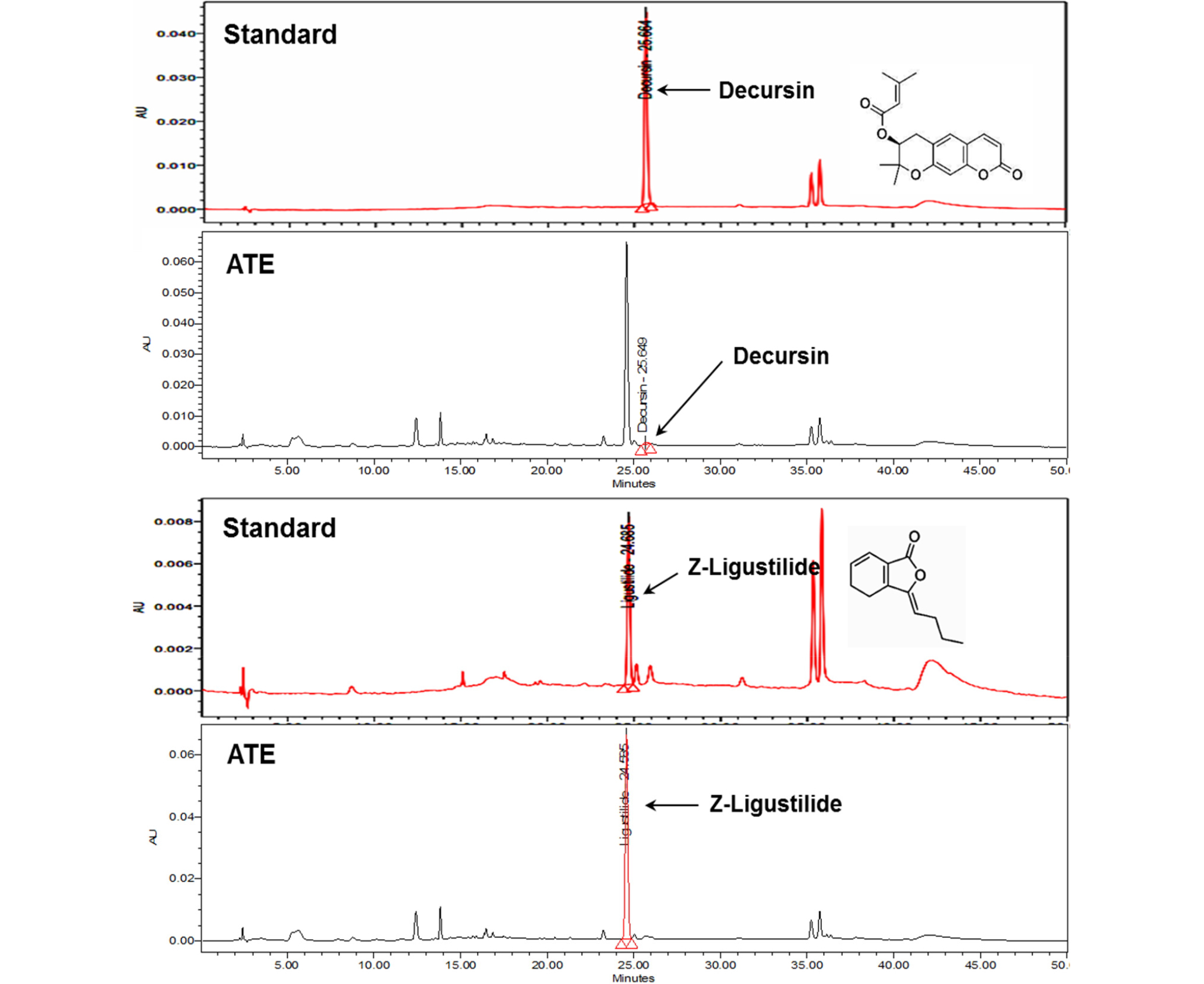
Determination of content of decursin and Z-ligustilide in ATE
Decursin and Z-ligustilide are reliable marker compounds for the authenticity of ATE(Nam et al., 2014). When the content of decursin and Z-ligustilide were determined in ATE by HPLC method, the retention times of decursin and Z-ligustilide were approximately 25.66 min and 24.68 min, respectively (Fig. 1). ATE contained 0.60 ㎍/㎎ (0.06%) of decursin and 84.34 ㎍/㎎ (8.43%) of Z-ligustilide (Fig. 1).
The content of total phenolic and total flavonoid
Total phenolic and total flavonoid content were expressed as a quercetin equivalents (QE) and gallic acid equivalents (GAE), respectively. ATE (1 ㎎) contained flavonoid as a 5.52 ㎍/QE and phenolic as a 237.27 ㎍/GAE (Table 1).
Table 1. Total phenolic and total flavonoid content of A. tenuissimaextract
| Total phenolic (z/㎎dried extract) | Total flavonoid (㎍QEy/㎎dried extract) | |
| Ethanol extract of A. tenuissimaroot (ATE) | 237.27 ± 13.24x | 5.52 ± 0.07x |
Effect of ATE on the cell viability
MTT assay was conducted at both 24 and at 48 h to determine the cytotoxicity of ATE in B16F10 cells. ATE up to 1000 ㎍/㎖ did not show significant cell death at both 24 (Fig. 2A) and 48 h (Fig. 2B). However, ATE at the concentration of 2000 ㎍/㎖ displayed approximately 40% reduction of cell viability at both 24 and 48 h compared to untreated control. Hence, subsequent experiments were conducted at below 1000 ㎍/㎖ of ATE.
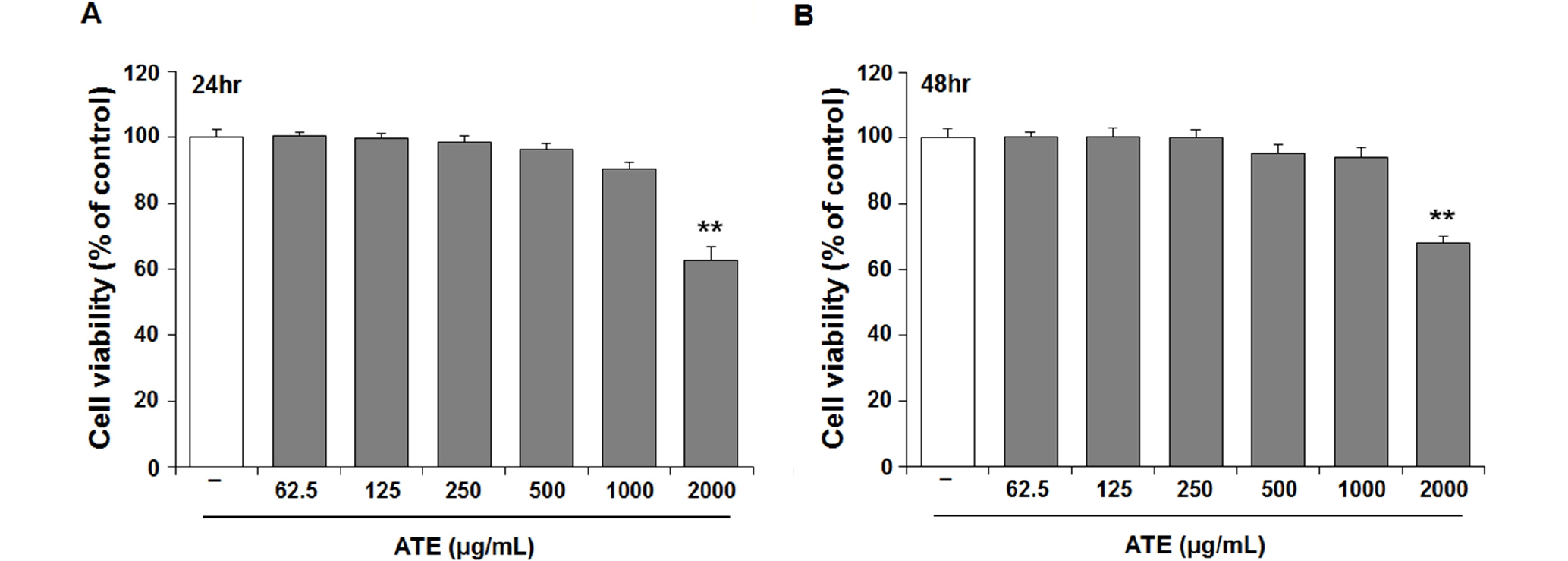
Fig. 2.
Effect of the Angelica tenuissima root extract (ATE) on cell viability in B16F10 melanocytes. B16F10 cells were treated with the ATE at the indicated concentrations. Cell viabilities were measured using MTT assay at (A) 24 h and (B) 48 h after treatment. The viability of untreated control cells was defined as 100%. The results are mean ± SEM of quintuplicates from a representative experiment (**p<0.01; significantly different from the control).
Antioxidant capacity of ATE
The radical scavenging activities of ATE were determined by DPPH and ABTS assays. As shown Fig. 3A and B, DPPH and ABTS scavenging activity were increased as the concentration of ATE increased. The effective concentration for 50% scavenging activity (IC50) was determined by the regression equation, and the IC50 values (㎎/㎖) of DPPH and ABTS radical scavenging activity were 2.78 and 0.76, respectively. The reducing power represents the degree of electron donating capacity and is determined by the ability of antioxidant to reduce Fe3+-ferricyanide complex to ferrous form (Fe2+). The reducing power of ATE was linearly proportional to the concentration and was found to reach 0.82±0.01 at 1000 ㎍/㎖ and have EC50 values of 0.56 ㎎/㎖ (Fig. 3C).
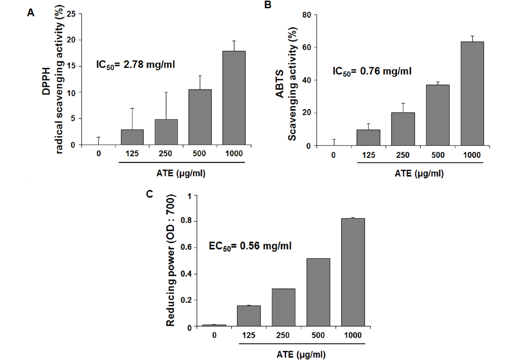
Fig. 3.
Radical scavenging activities of the Angelica tenuissima extract (ATE) in various concentrations. Antioxidant activities of ATE were determination by (A) DPPH radical scavenging activity, (B) ABTS radical scavenging assay, and (C) reducing activity power of ATE. Each value represents a mean ± SEM from triplicate determinations.
Inhibitory effect of ATE on the melanin production in B16F10 cells
Suppression of melanin production is one of key events required in skin whitening. Effect of ATE on the melanin production in B16F10 cells were determined in the presence or absence of α-MSH. Arbutin is widely used in a topical form for dermatologic and cosmetic uses and thus used as a positive control for determination of melanin production. As shown in Fig. 4A and 4B, melanin production was not significantly enhanced by ATE or arbutin in the absence of α-MSH stimulation (p>0.05). This result indicates that ATE at given dose did not affect melanin production in unstimulated condition. Following α-MSH stimulation, the melanin production was significantly increased (156.32±2.21% vs. untreated control, p<0.05). However, the α-MSH-stimulated increase of melanin production was significantly suppressed by ATE treatment with a dose-dependent mode (Fig. 4C and D). These results suggest that ATE can suppress α-MSH-mediated stimulation of melanin production.
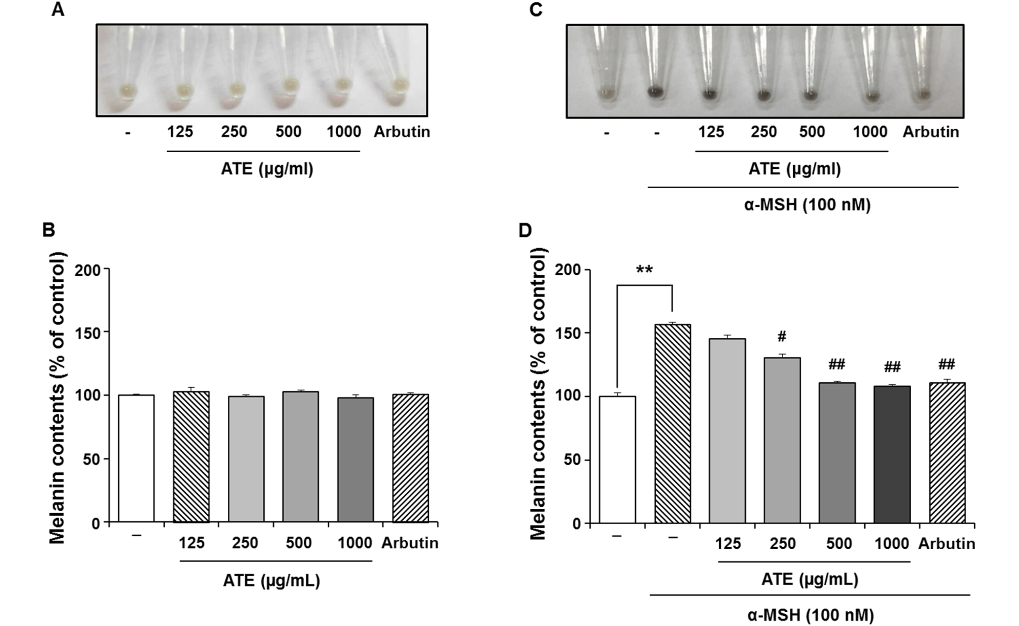
Fig. 4.
Regulatory effect of the Angelica tenuissima extract (ATE) on melanin production in the presence or absence of α-melanocyte stimulating hormone (α-MSH) insult. (A) Images of pellets of B16F10 cells harvested and (B) the determination of relative melanin contents in the absence of α-MSH after treatment of ATE (125-1000 ㎍/㎖) or arbutin (100 ㎍/㎖) for 48 h. (C) Images of pellets of B16F10 cells harvested and (D) the determination of relative melanin contents with α-MSH after treatment of ATE (125-1000 ㎍/㎖) or arbutin (100 ㎍/㎖) for 48 h. Data are the mean ± SEM and expressed as a percentage of untreated control. *p<0.05 and **p<0.01 vs. untreated control and #p<0.05, ##p<0.01 vs. α-MSH-treated control.
Effect of ATE on the in vitro tyrosinase and α-glucosidase activities
Tyrosinase has been used extensively as a target enzyme to search for whitening agents, because this enzyme is a rate- limiting enzyme in the process of melanogenesis (Bin et al., 2016). Arbutin behaves as a competitor inhibitor of L-tyrosine or L-dopa for tyrosinase and thus melanin production will be suppressed by arbutin (Maeda and Fukuda, 1996). As shown in Fig. 5A, arbutin (100 ㎍/㎖) caused 41.5% reduction of tyrosinase activity. ATE at the concentration of 125, 250, 500 and 1000 ㎍/㎖ also suppressed the tyrosinase activity ranging from 14.2 to 24.1% in α-MSH-stimulated B16F10 cells.
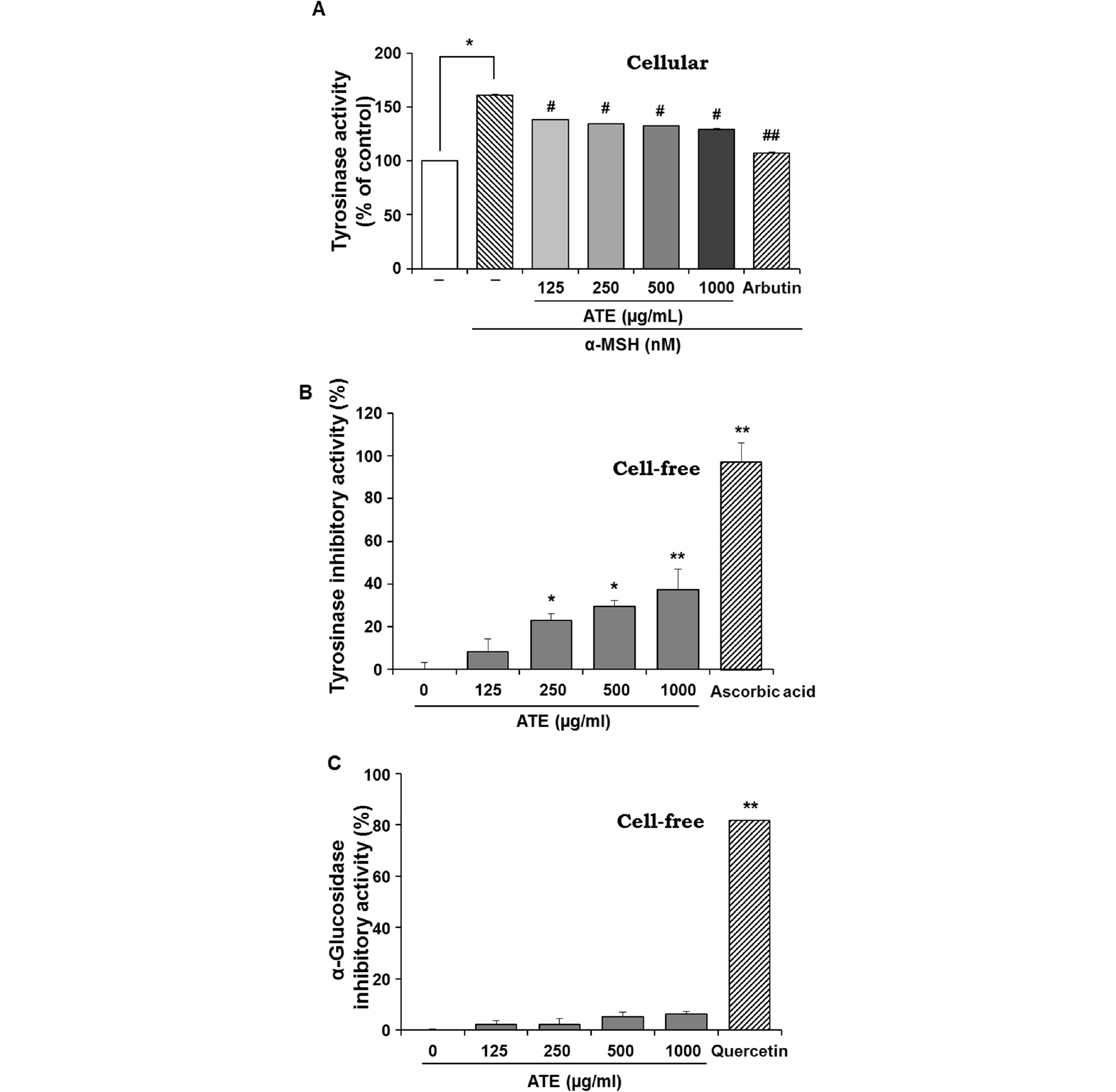
Fig. 5.
Effects of the Angelica tenuissima extract (ATE, 125- 1000 ㎍/㎖) on (A) B16F10 cellular and (B) a cell-free tyrosinase activities. Arbutin (100 ㎍/㎖) or ascorbic acid (50 ㎍/㎖) was used as a positive for tyrosinase activities in cellular or cell-free system, respectively. (C) α-Glucosidase inhibitory activities of ATE in a cell-free system. Quercetin (10 ㎍/㎖) was used as positive control of α-glucosidase inhibitor. Tyrosinase and α-glucosidase regulatory activities were determined by colorimetric method. Data are the mean ± SEM and expressed as a percentage of untreated control. *p<0.05, **p<0.01 vs. untreated control and #p<0.05, ##p<0.01 vs. α-MSH-treated control.
Change of tyrosinase activity in the cells treated with the herb extract is also a useful indication about its depigmentation activity to the cells. We further determined the tyrosinase inhibitory activity of ATE in cell-free system. Ascorbic acid potently inhibits tyrosinase through its interacting with four amino acid units in the active site of this enzyme in silico approach (Senol et al., 2014). Compared to ascorbic acid (50 ㎍/㎖) which complete blocks the tyrosinase activity, ATE at concentration of 250, 500 and 1000 ㎍/㎖ could significantly suppress tyrosinase activity with less potency than that of ascorbic acid (Fig. 5B).
N-glycan processing of tyrosinase is mediated with the aid of α-glucosidase in endoplasmic reticulum (Petrescu et al., 1997). It appears that reduction of melanin synthesis is strongly associated with enzymatic activity of tyrosinase and its N-glycan processing in cells. When α-glucosidase is inhibited, melanin synthesis is suppressed due to loss of enzymatic activity of tyrosinase and transport of tyrosinase to the melanosome (Bin et al., 2016). Unlike tyrosinase, the enzymatic activity of α-glucosidase, which can be strongly inhibited by quercetin (10 ㎍/㎖), was not significantly inhibited in the presence of ATE at a given dose (Fig. 5C). This result suggests that ATE does not contain remarkable amount of flavonoids involved in the inhibition of α-glucosidase as shown by quercetin. Based on the result of Fig. 5A and 5B, it is suggested that ATE has a significant inhibitory potential against tyrosinase activity in both cellular and cell- free system.
Discussion
In this study, the antioxidant and anti-melanogenic activity of 70% ethanol extract of A. tenuissima (ATE) was demonstrated either in vitro cell-free or in B16F10 cell system. Moreover, it was analysed and quantified using HPLC-PDA for two marker components, the pyranocoumarin compound, decursin and a naturally occurring phthalide, Z-ligustilide from the root of A.tenuissima.
It has become increasingly evident that oxidative stress would lead several skin diseases and skin aging, and antioxidants play protective role for skin health and regulation of the growth of melanocytes and melatonin (Kohen, 1999; Gašperlin and Gosenca, 2011; Peres et al., 2011). The present results showed that ATE presents the potent antioxidant activity in a concentration- dependent manner, which was measured by the method of DPPH, ABTS radical scavenging assay, and reducing power assay. There was reported that the total content of phenolic compounds and flavonoids are highly correlated with antioxidant properties of plant extracts (Komes et al., 2011). Moreover, many researches have focused on the biological activities of phenolics, due to their potent antioxidants, reducing agents and free radical scavengers (Kähkönen et al., 1999; Chandra et al., 2014). Our results showed that ATE contained phenolic as a 237.27 ㎍·GAE/㎎ and flavonoid as a 5.52 ㎍·QE/㎎, therefore, the antioxidant activity of ATE could be attributed to its phenolic compound and flavonoid contents.
White skin color is strongly associated with lessened production of melanin and its synthesis is mediated by tyrosinase in melanocytes. Hence, the identification of tyrosinase inhibitory potential of compound or extract has been widely used as the first line trial. Here, we found that ATE was able to inhibit in vitro mushroom tyrosinase with a dose dependent manner. Indeed, plant tyrosinase is different from mammalian tyrosinase because of its unique requirements for substrate and cofactor as well as its different sensitivity to inhibitors (Virador et al., 1999). Therefore, some plant extracts showing inhibitory activity to mushroom tyrosinase in vitro did not reduce pigmentation activity in cells (Zhong et al., 2006). In case of ATE, the suppressive effect of tyrosinase shown in vitro system was also reproduced in α-MSH stimulated B16F10 cells and resulted in lessened contents of melanin in the cells. α-Glucosidase activity is another determining factor involved in melanin synthesis because N-glycosylation of tyrosinase is necessary for its transporting into melanosome. ATE at a given dose did not reduce in vitro α-glucosidase activity. These results suggest that the anti-melanogenic capacity of ATE will be associated not with α-glucosidase inhibition but with inhibition of tyrosinase (Fig. 5).
As natural products are a rich reservoir for discovering new bioactive compounds because of their diversity and complexity structures. The present study suggests that ATE has a strong possibility of multi-target strategy with different mechanisms involved in skin whitening. Therefore, ATE may be applicable combined with other established skin whitening compounds.
