

Introduction
Materials and Methods
Experimental material
Experimental material extraction
Fraction preparation
Isolation of complex saponin
Cell culture
Lactate dehydrogenase (LDH) assay
Nitric oxide (NO) assay
Prostaglandin E2 (PGE2) assay
Statistical analysis
Results
Cytotoxicity assay
NO production inhibitory effect
PGE2 production inhibitory effect
Discussion
Introduction
Drinking and smoking environmentally toxic compounds and radioactive substances, which are increasing due to modern industrialization, cause expression of reactive oxygen species (ROS) and increase oxidative stress in the body (Kim et al., 2012a). Depending on the kind of ROS, they can include oxygen-oriented radicals (O2–), •OH and non-radical species (H2O2 and O2), as well as ROOH, ROO•, RO•, and HOCl, which can be expressed by the reactions of ROS with other biocomponents. In the body, ROS cause DNA fragmentation, peroxidation, and protein inactivation, thereby inhibiting biological functions and generating various pathogenic factors. Physiological actions to inhibit ROS include inhibition of oxidative ROS by donating electrons, and superoxide dismutase (SOD)-like activity to convert the super\oxide anion radical into normal oxygen. Accordingly, interest in obtaining compounds exhibiting physiological activities, such as antioxidant activity, from food groups easily accessible to modern people is increasing (Kang et al., 1996; Kim et al., 2020; Lee et al., 2012; Ryu et al., 2012).
In response to harmful substances, such as external physical and chemical stimuli, viruses, bacteria, and bacteria that penetrate into the body, inflammatory reactions due to free radicals occur and secrete various inflammatory mediators to restore damaged tissues and preserve bodily functions. However, if the inflammatory response continues, excessive secretion of inflammatory mediators leads to cancer and increases insulin resistance, which acts as a mediator of various diseases, such as diabetes (Cheon et al., 2009; Nishida et al., 2007). The main cells involved in inflammation are macrophages, which are activated by various stimuli or cytokines secreted by immune cells to produce inflammatory cytokines, nitric oxide (NO) and prostaglandin E2 (PGE2), resulting in edema and pain. The inflammatory responses include fever and erythema and activate migration of immune cells to inflammation-inducing tissues (Kim et al., 2012c). Among these responses is the production of NO, which is highly reactive, from L-arginine by NO synthase (NOS). Both constitutive NOS and inducible NOS (iNOS) are produced. In particular, iNOS reportedly is expressed in various cells, such as smooth-muscle cells, hepatocytes, bone-marrow cells, macrophages, and monocytes, when stimulated by external stress or inflammatory cytokines to produce a large amount of NO. Consequently, iNOS, COX-2, NO, and PGE2 are representative inflammation-inducing factors of immune cells. On the other hand, as stated in the bulletin, one of the other causes of the inflammatory response is oxidative stress in the body. In particular, drinking, smoking, and obesity are known to increase oxidative ROS and cause acute and chronic inflammation. Therefore, intake of ingredients with excellent antioxidant effects can suppress the inflammatory response by reducing oxidative stress (Bak et al., 2009; Kim et al., 2012b; Lee et al., 2012; Uttara et al., 2009).
Recently, there has been increasing demand for functional health food because it is easily exposed to lifestyle-related and degenerative diseases due to changes in the westernized diet (Cho et al., 2014). Research that investigates various natural plant components as functional materials is being attempted, which requires analysis of the physiologically active components of various resource plants and use in various fields (Hwang et al., 2010; Lee et al., 2014). One study of the complex saponins of Pueraria lobata flower and Phaseolus angularis reported representative analysis results for the compounds, but studies using pathogenic factors on the physiological activities of these saponins are insufficient. Therefore, the aim of the present study was to treat RAW 264.7 macrophages induced by lipopolysaccharide (LPS) stimulation with the complex saponins, P. lobata flower and P. angularis, to evaluate their anti-inflammatory activities by NO and PGE2 assay and their potential use as functional foods.
Materials and Methods
Experimental material
The P. lobata flower and P. angularis used in this experiment were purchased at Gyeongdong Market in Jegi-dong, Dongdaemun-gu, Seoul in January 2020 and were identified by Dr. J. N. Lee. The extraction sample was used after being dried in the shade and then pulverized. A voucher specimen (NIHA.P-26-2) was deposited in the herbarium of the Highland Agriculture Research Institute, Pyeongchang, Korea.
Experimental material extraction
P. lobata flower and P. angularis were pulverized into a powder weighing 3 ㎏ at a 5:5 mixing ratio and then chilled with ethanol solvent at room temperature for 5 months. After filtering the ethanol extract with qualitative filter paper, the filtrate was concentrated on a rotary vacuum evaporator (Tokyo Rikakikai Co., Tokyo, Japan) to obtain an ethanol extract. Table 1 lists the measurement results.
Table 1.
Yield of the Pueraria lobata flower and Phaseolus angularis ethanol extract.
| Sample | Ethanol extractz | |
| Weight (g) | Yield (%) | |
| P. lobata flower and P. angularis | 250 | 8.3 |
Fraction preparation
After suspending 250 g of the P. lobata flower and P. angularis ethanol extract in 0.8 L of distilled water, n-hexane (1 L × 3), chloroform (1 L × 3), ethyl acetate (1 L × 3), and butyl alcohol (BuOH) were examined in the order of polar to nonpolar solvents. Table 2 lists the measurement results after eluting with 1 L × 3 and vacuum concentration of each fraction.
Table 2.
Yield of P. lobata flower and P. angularis organic solvent fractions.
| Solvent fractionz | P. lobata flower and P. angularis | ||
| Weight (g) | Yield (%) | ||
| n-Hexane | 34.1 | 13.7 | |
| Chloroform | 48.3 | 19.3 | |
| Ethyl acetate | 57.3 | 22.9 | |
| Butyl alcohol | 60.4 | 24.2 | |
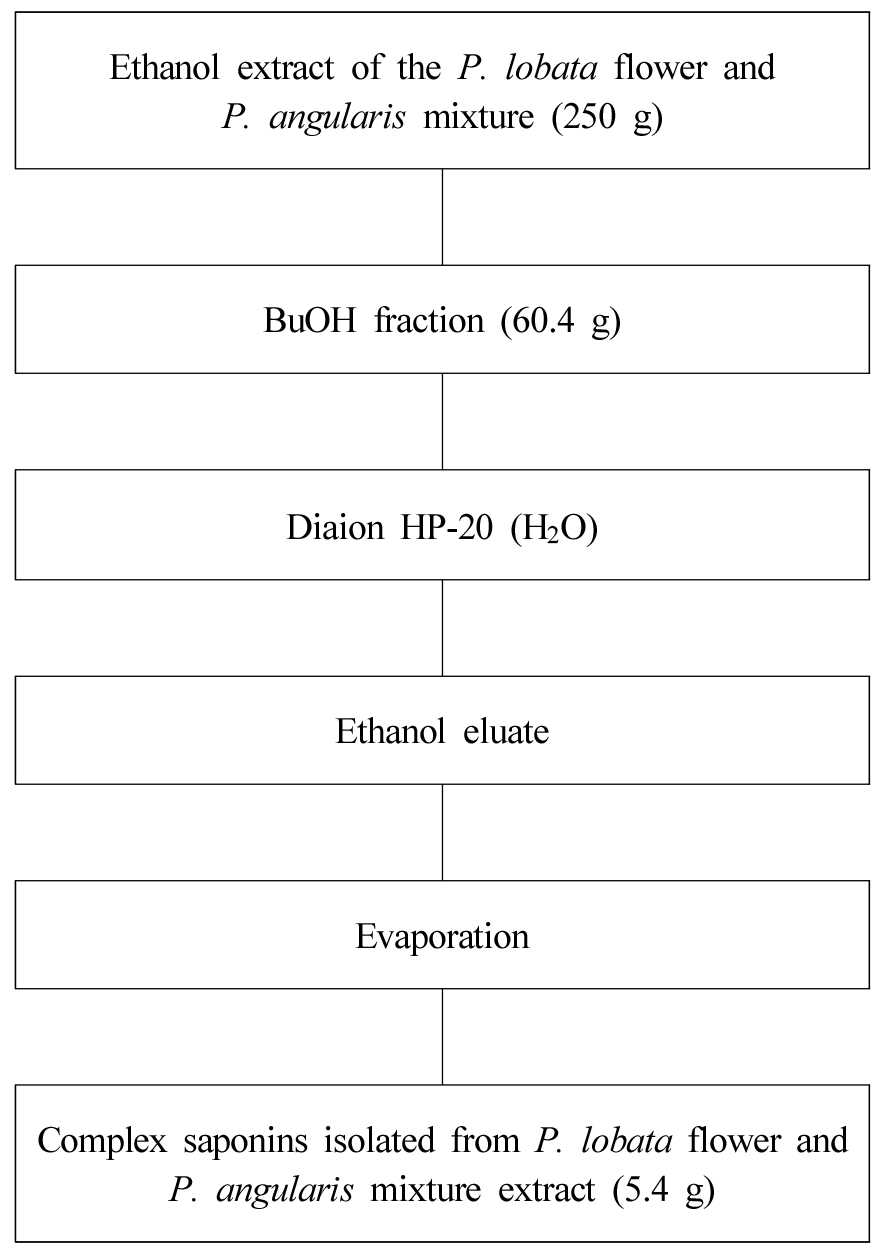
Isolation of complex saponin
Dried P. lobata flower and P. angularis were cut and extracted three times with ethanol under reflux and evaporated to obtain a viscous mass. The ethanol extract was dried under vacuum. The P. lobata flower and P. angularis mixed ethanol extracts were fractionated with butyl alcohol (BuOH). The Diaion HP-20 resin adsorption method was used to isolate the complex saponins from the BuOH fraction(Nam, 2022). The isolate complex saponins (5.4 g) were used as the sample for the vascular relaxation experiment (Fig. 1).
Cell culture
The RAW 264.7 cell line was cultured in a Dulbecco’s Modified Eagle Medium (DMEM) culture medium supplemented with 10% fetal bovine serum (FBS), penicillin 10 unit/mL, streptomycin 100 μg/mL, and 2 mM L-glutamine at 37℃ in a 5% CO2–95% air incubator. After washing the monolayer cultured cells in a culture flask twice with the basal medium, the cells were detached by treatment with a 2.5% trypsin solution, and the suspended cells were neutralized with a serum medium and centrifuged, then washed once. After measuring the number of cells using a hemocytometer, adjusting the number of cells to a concentration of 1×10⁴ cells/mL, putting 500 μL of each into a 24-well plate, and culturing for 24 hours.
Lactate dehydrogenase (LDH) assay
To check the degree of toxicity of cells (cell viability), the LDH test method (Sigma Chemical Co., USA) was used. In a 96-well plate (Falcon T.M., USA), the cells were equally distributed as 1 µ105 cells/mL, and the medium was removed after incubation for 24 hours. After washing with phosphate-buffered saline (PBS) (Bioneer Co., Korea) buffer, a single 100 μL sample of H2O2 (BBC Biochemical, USA) and multiple 100 μL samples of various concentrations were added to each well and incubated for 45 minutes. Then, the supernatant was removed and after washing twice with PBS, 200 μL of medium was injected and incubated for 24 hours. Then, 50 μL of 5 ㎎/mL LDH reagent was added to the culture medium, incubated for 4 hours, and the supernatant was removed followed by addition of 100 μL of dimethyl sulfoxide (Sigma Chemical Co., USA). An enzyme-linked immunosorbent assay (ELISA) microplate reader (BioTek Instruments Co., USA; wavelength = 540 ㎚) was used to measure absorbance and calculate viability.
Nitric oxide (NO) assay
Expression of NO generated from RAW 264.7 macrophages using Griess reagent solution [1% (w/v) sulfanilamide in 5% (v/v) phosphoric acid and 0.1% (w/v) naphthyl ethylene diamine-HCl] The amount was measured in the form of NO2− present in the cell culture medium. Then, 100 μL of Griess reagent solution was mixed and reacted in a 96-well plate for 10 minutes, and the absorbance was then measured at a wavelength of 540 ㎚ by an ELISA microplate reader (BioTek Instruments Co., USA).
Prostaglandin E2 (PGE2) assay
After taking the sample-treated cell culture, the expression level of PGE2 was measured according to the kit manual using the prostaglandin assay design kit (Amersham Pharmacia Biotech Co., USA). RAW 264.7 macrophages were aliquoted into a 24-well plate at a unit concentration of 1×10⁵ cells/mL, treated with each concentration, and cultured for 24 hours, followed by addition of LPS (100 ng/mL) and incubated for another 24 hours. After incubation, the cell culture supernatant was taken and tested. After adding 200 μL of Ellman’s reagent solution and reacting for 60 to 90 minutes, the absorbance was measured on the ELISA microplate reader (BioTek Instruments Co., USA) at a wavelength of 420 ㎚.
Statistical analysis
After three repeated independent experiments, the experimental value was expressed as the inhibition rate (%). The significance of statistical analysis was assessed by performing Student’s t-test, which tests the null hypothesis (H0) that the mean value is equal to a specific value μ0 for the normal population and corresponds to the case in which the standard deviation δ is unknown.
Results
Cytotoxicity assay
Prior to examining the inhibitory effects of NO and PGE2 production on the complex saponins isolated from P. lobata flower and P. angularis, to set the treatment capacity, we performed an experiment to identify the optimum treatment dose that would exclude the possibility of an inhibitory effect of the inflammatory mediators due to toxicity to RAW 264.7 macrophages; that is, the dose at which toxicity was not shown in the LDH assay. The concentration at which the minimum cell viability was ≥90% was judged to be the optimal treatment dose. As can be seen in Table 3, The complex saponins isolated from P. lobata flower and P. angularis exhibited cytotoxicity at all treatment concentrations of ≤100 μg/mL. We confirmed that the survival rate was not shown.
Table 3.
Complex saponin isolated from P. lobata flower and P. angularis on the cell viability of RAW 264.7 cells.
| LDH assayz | |
| Complex saponins isolated from P. lobata flower and P. angularis (㎍/mL) | Cell viability (%) |
| 0 | 100 |
| 12.5 | 100 |
| 25 | 99 |
| 50 | 95.2 |
| 100 | 90.1 |
zCells were treated with 0, 12.5, 25, 50, and 100 ㎍/mL of the complex saponins isolated from P. lobata flower and P. angularis for 24 hours. The amount of viable cells was determined by LDH assay. There were no significant differences between the non-treated group and complex saponins isolated from the P. lobata flower - and P. angularis-treated groups. Data are expressed as the suppression rate (%).
NO production inhibitory effect
To determine if the complex saponins isolated from P. lobata flower and P. angularis significantly inhibited NO production in LPS-stimulated RAW 264.7 macrophages at 0, 12.5, 25, and 50 μg/mL, we first measured NO by treatment with RAW 264.7 macrophages at a concentration of 100 μg/mL. We found that NO production was reduced by about 62.2% in a sample at a concentration of 50 μg/mL, and NO production was approximately 73.4% in a sample with a concentration of 100 μg/mL suppressed. The complex saponins isolated from P. lobata flower and P. angularis were used to treat RAW 264.7 macrophages at different concentrations to confirm the effect on NO production. As shown in Table 4, NO production was inhibited in a concentration-dependent manner. Therefore, the complex saponins were judged to have a significant effect on the inhibition of other inflammatory metabolites or mediators, so an additional physiological activity experiment was conducted.
Table 4.
Complex saponins isolated from P. lobata flower and P. angularis on NO production in LPS-stimulated RAW 264.7 cells.
| NO Inhibition Assayz | |
| Complex saponins isolated from P. lobata flower and P. angularis (㎍/mL) | NO Inhibition (%) |
| 0 | N.D.y |
| 12.5 | N.D. |
| 25 | N.D. |
| 50 | 62.2 |
| 100 | 73.4 |
zNO concentration was measured by use of Griess reagent after RAW 264.7 cells were treated with complex saponins isolated from P. lobata flower and P. angularis for 24 hours. The percent inhibition values of NO production were calculated relevant to the LPS treatment as 100% and the control treatment as 0%. The data are expressed as the suppression rate (%),
PGE2 production inhibitory effect
The complex saponins isolated from P. lobata flower and P. angularis, which showed a significant inhibitory effect in the NO screening experiment, was measured for each concentration by concentration. The complex saponins isolated from P. lobata flower and P. angularis were used to treat RAW 264.7 macrophages at concentrations of 0, 12.5, 25, 50, and 100 ㎍/mL to measure the effect of inhibiting PGE2 production. We found that PGE2 production was inhibited by about 60.4% in the sample and by about 71.0% in the 100 μg/mL sample. We confirmed that the concentration-dependent inhibition of PGE2 production was increased by LPS stimulation (Table 5). In the previous NO assay, the complex saponins isolated from P. lobata flower and P. angularis showed the highest inhibitory effect at a concentration of 100 μg/mL, and in the PGE2 assay, they also showed the highest inhibitory effect at 100 μg/mL. Increasing concentration effectively inhibited PGE2 production. The complex saponins isolated from P. lobata flower and P. angularis were used to confirm the inhibitory effect on the production of NO and PGE2, an inflammatory mediator induced by LPS stimulation in RAW 264.7 macrophages, which are murine macrophage cells. Through this approach, we attempted to select the optimal treatment concentration in the future anti-inflammatory effect confirmation experiment. As a result of screening according to the treatment concentration set by LDH assay, the complex saponins isolated from P. lobata flower and P. angularis showed strong effects in the NO and PGE2 production inhibition tests. Therefore, the complex saponins isolated from P. lobata flower and P. angularis were judged to be suitable for use in future studies on the pharmacological activity in different diseases.
Table 5.
Inhibitory effects of the complex saponins isolated from P. lobata flower and P. angularis on PGE2 production in LPS-stimulated RAW 234 cells.
| PGE2 Inhibition Assayz | |
|
Complex saponins isolated from P. lobata flower and P. angularis (㎍/mL) |
PGE2 Inhibition (%) |
| 0 | N.D.y |
| 12.5 | N.D. |
| 25 | N.D. |
| 50 | 60.4 |
| 100 | 71.0 |
zPGE2 production was measured by use of PGE assay kits after RAW 264.7 cells were treated with the complex saponins isolated from P. lobata flower and P. angularis for 24 hours. The inhibition percentages of PGE2 production were calculated relative to LPS treatment, taken as 100%, and the control treatment, taken as 0%. Data are expressed as the suppression rate (%),
Macrophages in the body produce and secrete secondary mediators, such as inflammatory cytokines, NO, PGE, and leukotrienes. These inflammatory mediators have a very important role in regulating innate and acquired immunity (Iontcheva et al., 2004; Yun et al., 2010). However, when these substances are secreted in excess, they can cause Crohn’s disease, a type of chronic inflammatory bowel disease, rheumatoid arthritis, and autoimmune diseases (Hilliquin et al., 1997; Nava and Moncada 1992: Yang et al., 2019). The present study investigated the inhibitory effect on inflammatory mediators of LPS-induced RAW 264.7 macrophages by the complex saponins isolated from P. lobata flower and P. angularis. These saponins were subjected to an LDH assay to assess the degree of cytotoxicity (cell viability), and to set an effective concentration range that does not show toxicity. The effective concentration range of the complex saponins isolated from P. lobata flower and P. angularis was from 0 to 100 μg/mL, as shown by testing at the individual concentrations of 0, 12.5, 25, 50, and 100 μg/mL. Endotoxin, also referred to as LPS, was treated to induce the production of NO, an inflammatory index, and each fraction was treated to confirm the inhibitory effect on NO production, and the inhibitory effect on PGE2-production also was confirmed. The complex saponins isolated from P. lobata flower and P. angularis were confirmed to exhibit significant anti-inflammatory activity by inhibiting the production of NO and PGE2 in a concentration-dependent manner. Since P. lobata flower and P. angularis are legumes, it is expected that the key ingredients showing physiological activity are triterpenoid saponins.
Discussion
Inflammation is a biological response to infection and damage to specific tissues in the body, and the main mediators are immune cells. Inflammation is divided into acute and chronic inflammation, and can be classified according to the size of the infection site and bodily tissue damage. When the extent of inflammation is greatly expressed or does not proceed in the form of acute inflammation, it progresses to chronic inflammation or arthritis. At a low level, obesity can also be classified as an inflammatory disease. In the past, legumes were mainly used as folk recipes to treat inflammation, so the complex saponins isolated from P. lobata flower and P. angularis were used to evaluate cytotoxicity and anti-inflammatory activity. The inhibitory effects of inflammatory mediators, such as NO and PGE2, were confirmed after stimulation with LPS, an inflammation-inducing factor, in RAW 264.7 macrophage cells. When the inhibition rate (%) of the inflammatory mediators was measured after treatment with the complex saponins isolated from P. lobata flower and P. angularis, the concentration that significantly inhibited the production of NO and PGE2 was 100 μg/mL. We confirmed that NO and PGE2 expressions were both inhibited by ≥50% at 100 μg/mL. Therefore, the study results showed that the complex saponins isolated from P. lobata flower and P. angularis exhibited significant anti-inflammatory effects and that these compounds potentially can be used therapeutically. An additional use would be as functional health food since P. lobata flower and P. angularis are legumes that can be eaten. Additional studies are needed on the inhibitory mechanisms of IL-4 and IL-13, which are cytokines related to inflammation, and on iNOS and COX-2, which are inflammatory marker proteins, that are key components involved in anti-inflammatory activity.
