

서 언
재료 및 방법
실험재료
추출물 제조
RAW264.7 세포배양
Nitric oxide (NO) 측정
SDS-PAGE 및 western blot 분석
Reverse transcriptase-polymerase chain reaction (RT-PCR)
통계분석
결과 및 고찰
망개나무 추출물의 NO 생성 억제능
iNOS, COX-2, IL-1β, TNF-α 및 IL-6 발현 억제
망개나무 잎 추출물의 NF-κB의 활성 억제 효과
망개나무 잎 추출물의 MAPK의 활성 억제 효과
적 요
서 언
염증이란 여러 자극에 의해 손상된 조직을 복구 및 재생하기 위해 일어나는 필수적인 생체 내 방어기전으로 다양한 병리 및 생리학적인 반응에 중요한 역할을 한다(Lee et al., 2006, Wang et al., 2018). 염증인자들은 염증성 질환을 일으키는 것만 아니라 주변 조직의 손상과 함께 생체기능에 이상 또는 심각하게는 관절염, 퇴행성 뇌질환, 암과 같은 질병으로 이어져 만성질환을 유발하기도 한다(Atreya and Neurath, 2005; Rabson et al., 2005). 염증이 발현되는 신호경로는 nuclear factor-κB (NF-κB)와 c-Jun NH2-terminal kinase (JNK), extracellular signal- regulated kinase (ERK), p38을 포함한 mitogen activated protein kinase (MAPK)가 중요한 역할을 한다(Guha et al., 2001). 현재 사용되고 있는 대부분의 항염증제는 장기로 복용할 경우 혈압상승, 신장기능 저하, 위장관 궤양 등의 부작용과 함께 혈전형성, 심근경색 등 순환계 질환도 유발할 수 있다고 보고되었다(Boumpas et al., 1993). 이러한 이유 때문에 최근에는 부작용이 적으면서 자연에 서식하고 있는 천연물 소재를 이용하여 여러 질환의 치료제 개발에 관심이 높아지고 있는 추세이다.
망개나무[Berchemia berchemiaefolia (Makino) Koidz.]는 갈매나무목(Rhamnales) 갈매나무과(Rhammaceae) 먹넌출속(Berchemia)의 낙엽교목으로 국내에서는 1935년 충북 속리산 계곡에서 처음 발견되었으며, 일본에서도 비교적 넓게 분포하는 것으로 알려져 있다(Chang et al., 2005; Lee et al., 2005). Chi (1998)는 망개나무 뿌리가 중국에서 암 치료에 시험적으로 쓰이고 있는 식물자원으로 언급한 바 있으며, 염증 억제에 대한 효능을 보고하였고, 망개나무 뿌리 추출물의 항비만 및 비알코올성 지방간에 미치는 효과와 잎, 가지, 열매 등 부위별 추출물의 항산화 활성 및 면역조절에 관한 연구가 보고되어 있지만(Choi et al., 2015; Eo et al., 2020; Jin et al., 2014), 분자세포생물학적 작용기전에 대한 연구는 미비한 실정이다. 따라서 본 연구에서는 부작용의 우려가 적은 천연 유래 소재인 망개나무 추출물의 항염증 활성 및 작용기전을 구명하고자 한다.
재료 및 방법
실험재료
마우스 대식세포 RAW264.7 배양을 위해 사용된 배지인(DMEM)/F-12 1 : 1 +2.50 mM L-Glutamine, + 15mM HEPES buffer medium (DMEM/F-12)는 Lonza (Hyclone, USA)에서 구매하였다. Lipopolysaccharide (LPS)는 Sigma Aldrich (St. Louis. MO, USA)에서 구매되었으며, western blot의 분석을 위한 항체 IκB-α, p-ERK1/2, total-ERK1/2, p-p38, total- p38, p-JNK, total-JNK 및 β-actin은 Cell Signaling Technology (Danvers, MA, USA)에서 구매하였다.
추출물 제조
본 연구에서 사용된 시료인 망개나무는 2018년 8월에 괴산에서 수집하였으며, 확증표본은 국립산림과학원 산림약용자원연구소에서 보관하고 있다(표본번호: FMCBrGS-1808-1-3). 수집된 망개나무는 부위별(잎, 가지, 열매)로 분리 후, 동결 건조하여 각 시료 20 g에 70% 에탄올 400 mL 가하여 상온에서 48시간 교반하여 추출하였다. 추출 후 filter paper (No. 2, Whatman Co., Maidstone, England)에서 여과하였으며, 40℃ 이하의 중탕에서 감압 환류 냉각장치 (EYELA, Tokyo, Japan)로 농축하고 동결건조를 실시하였다. 완성된 시료 추출물[망개나무 잎 추출물(BBK-L), 가지 추출물(BBK-B), 열매 추출물(BBK-F)]은 dimethyl sulfoxide (DMSO)에 용해하여 실험에 사용하였다.
RAW264.7 세포배양
본 연구에서 사용된 마우스 대식세포 RAW264.7은 한국세포주은행에서 분양받아 실험에 사용하였다. RAW264.7은 10% fatal bovineserum (FBS)를 첨가한 100 U/mL penicillin 100 ㎍/mL streptomycin이 포함된 DMEM/F-12 배지로 5% CO2를 함유한 37% 배양기(Thermo, Germany) 에서 배양하였다. 세포는 2 ~ 3일에 한 번 계대하여 사용하였다.
Nitric oxide (NO) 측정
NO 측정은 Namkoong et al., 2015의 방법을 응용하여 측정하였다. 마우스 대식세포 RAW264.7을 12 well plate에 분주하고 24시간 동안 배양하였다. 24시간 후 망개나무 추출물을 0, 12.5, 25, 50 ㎍/mL 농도로 처리하고 6시간 동안 배양하였다. 6 시간 후 LPS를 1 ㎍/mL의 농도로 처리하고 18시간 배양하였다. Nitric oxide의 측정은 Griess reagent system (Sigma- Aldrich, St. Louis, MO, USA)을 이용하여 측정하였다.
SDS-PAGE 및 western blot 분석
망개나무 샘플이 처리된 세포에서 단백질을 추출하기 위해서 세포를 1 × phosphate-buffered saline (PBS)로 2회 세척한 후, radioimmunoprecipitation assay (RIPA) buffer (Boston Bio Products, Ashland, MA, USA)에 protease inhibitor cocktail (Sigma-Aldrich Co., St. Louis,MO, USA)와 phosphatase inhibitor cocktail (Sigma-Aldrich Co., St. Louis, MO, USA)를 포함하여 lysis시켜 단백질을 얻었다. Bicinchoninic acid (BCA) protein assay (Pierce Biotechnology Inc., Waltham, MA, USA)로 단백질 정량 후, 동일량의 단백질을 10% SDS-acrylamide에 loading하고 NC membrane (GE Helthcare life science, Germany)에 이동시킨 후 5% non-fat dry milk로 상온에서 1시간 동안 blocking하였다. 1시간 후, 1차 항체를 5% non-fat milk에 용해 후 4℃에서 14시간 동안 반응시키고 membrane을 0.05% tween-20이 포함된 tris-buffered saline (1 × TBS-T)으로 5분간 3회씩 세척하였다. 그 후 membrane에 2차 항체를 5% non-fat milk에 용해하여 1시간 처리하였고, TBS-T로 5분간 3회씩 세척 한 후 ECL western blotting substrate (Amersham Biosciences Co., Little Chalfont, England)를 이용하여 단백질을 확인하였다.
Reverse transcriptase-polymerase chain reaction (RT-PCR)
Total RNA 추출은 망개나무 잎(BBK-L)을 처리한 마우스 대식세포 RAW264.7에 RNeasy Mini kit (QIAGEN GmbH., Hilden,Germany)를 이용하여 수행하였으며, cDNA는 1 ㎍의 total RNA를 Verso cDNA kit (Thermo Fisher Scientific Inc., Waltham, MA, USA)를 이용하여 제조되었다. PCR은 PCR master mix kit (Promega Co., Madison, WI, USA)를 이용하여 수행하였고, 사용된 primer는 Table 1과 같다. PCR을 통하여 만들어진 DNA의 양을 확인하기 위해 1% agarose gel에 Safe shine green으로 염색하고 loading하여 전기영동으로 분리하였다. 이를 gel documentory system으로 확인(Biorad, Chemidoc, MP imaging system, USA)하였으며, housekeeping 유전자인 glyceraldehyde-3-phosphate dehydrogenase (GAPDH) 유전자를 포함하여 internal control로 사용하였다.
Table 1
Sequence of oligonucleotide primers used for RT-PCR
통계분석
모든 결과는 3 회 반복 측정 후 평균 ± 표준편차로 나타내었고, 처리간 유의성은 Student’s t-test로 검증하여 p-value 값이 0.05 미만일 때 통계적으로 유의하다고 판정하였다(Microsoft Exel 2010, Microsoft, Redmond, WA, USA).
결과 및 고찰
망개나무 추출물의 NO 생성 억제능
망개나무 추출물의 항염증 활성을 확인하기 위해 잎(BKK-L), 가지(BBK-B), 열매(BBK-F) 등 부위별 추출물의 NO 생성 억제능(%)을 측정한 결과, BBK-L에서 가장 높은 NO 생성 억제 효과를 나타냈다(Fig.1A). 따라서, 활성이 가장 높았던 BBK-L 추출물을 시료로 사용하였고 농도별(0, 12.5, 25, 50 ㎍/mL)로 처리하였을 때 농도가 높아짐에 따라 NO의 생성이 감소하는 것을 확인할 수 있었다(Fig. 1B). 또한 BBK-L 추출물의 처리 농도에서 세포독성을 나타내지 않는 것을 확인할 수 있었다(Fig. 1C). 대식세포는 면역체계에 있어 면역기능과 염증반응을 조절하는 중요한 역할을 담당하고 있으며, 과도한 NO의 증가는 염증성 질환을 발생시키며, 세포와 조직에 산화적 손상을 일으켜 유전자 변이, 신경 손상 등을 초래하기 때문에 NO 생성의 억제는 염증 관련 질환의 발병을 저해할 수 있는 방법으로 알려져 있다(Sim et al., 2017). 이러한 연구 결과를 통해 BBK-L 추출물은 농도 의존적으로 과도한 NO의 생성을 억제함으로써 염증반응을 조절한다고 보인다.
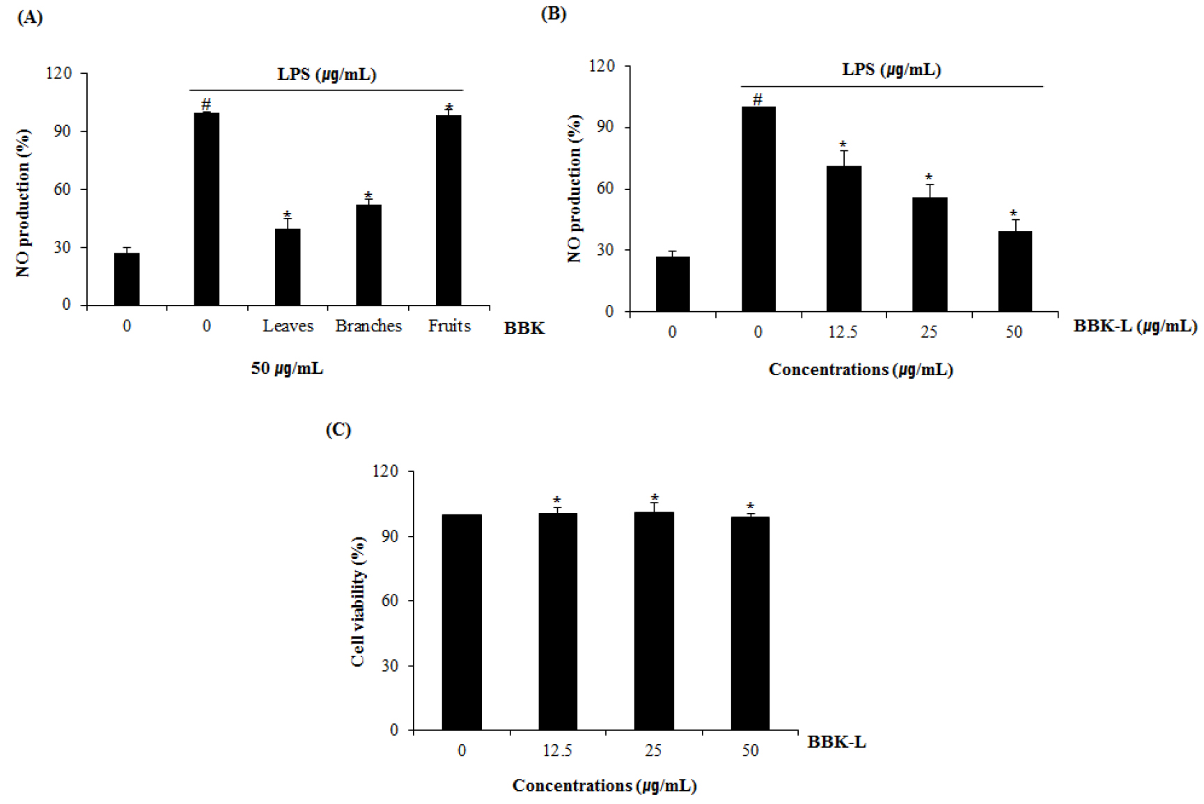
Fig. 1
Inhibitory effect of the extracts from leaves (BBK-L), branches (BBK-B) and fruits (BBK-F) of BBK against NO production in LPS-stimulated RAW264.7 cells. (A) RAW264.7 cells were pretreated with BBK-L, BBK-B and BBK-F for 6 hours and then co-treated with LPS (1 ㎍/mL) for 18 hours. (B) RAW264.7 cells were pretreated with BBK-L (0, 12.5, 25 and 50 ㎍/mL) for 6 hours and then co-treated with LPS (1 ㎍/mL) for 18 hours. NO production was measured by Griess assay. (C) RAW264.7 cells were treated with BBK-L for 24 h. Cell viability was measured using MTT assay. #p < 0.05 compared to the cells without the treatment, and *p < 0.05 compared to the cells treated with LPS alone.
iNOS, COX-2, IL-1β, TNF-α 및 IL-6 발현 억제
BBK-L 추출물이 iNOS, COX-2, TNF-α, IL-1β 및 IL-6 의 생성에 미치는 영향을 평가한 결과(Fig. 2), BBK-L 추출물이 처리되지 않은 세포에서는 LPS에 의해 iNOS, COX-2 TNF-α, IL-1β 및 IL-6에서 mRNA의 발현이 증가 되었지만, BBK-L의 농도가 증가할수록 mRNA 발현을 유의성 있게 억제하였다. 대식세포인 RAW264.7에 LPS로 자극을 주면 iNOS가 발현되면서 다량의 NO를 생성하여 염증반응을 일으키고, TNF-α, IL-1β 및 IL-6와 같은 전 염증성 cytokine의 발현이 과도하게 유도되어 COX-2 유전자의 발현을 자극하여 prostaglandin2 (PGE2) 등의 염증인자들을 생성시킨다고 알려져 있다(Guzik et al., 2003; Shin et al., 2012). 이러한 연구 결과는 BBK-L 추출물이 iNOS, COX-2, TNF-α, IL-1β 및 IL-6의 mRNA 발현 억제를 통하여 과도한 면역반응을 조절하는 것으로 나타났다.
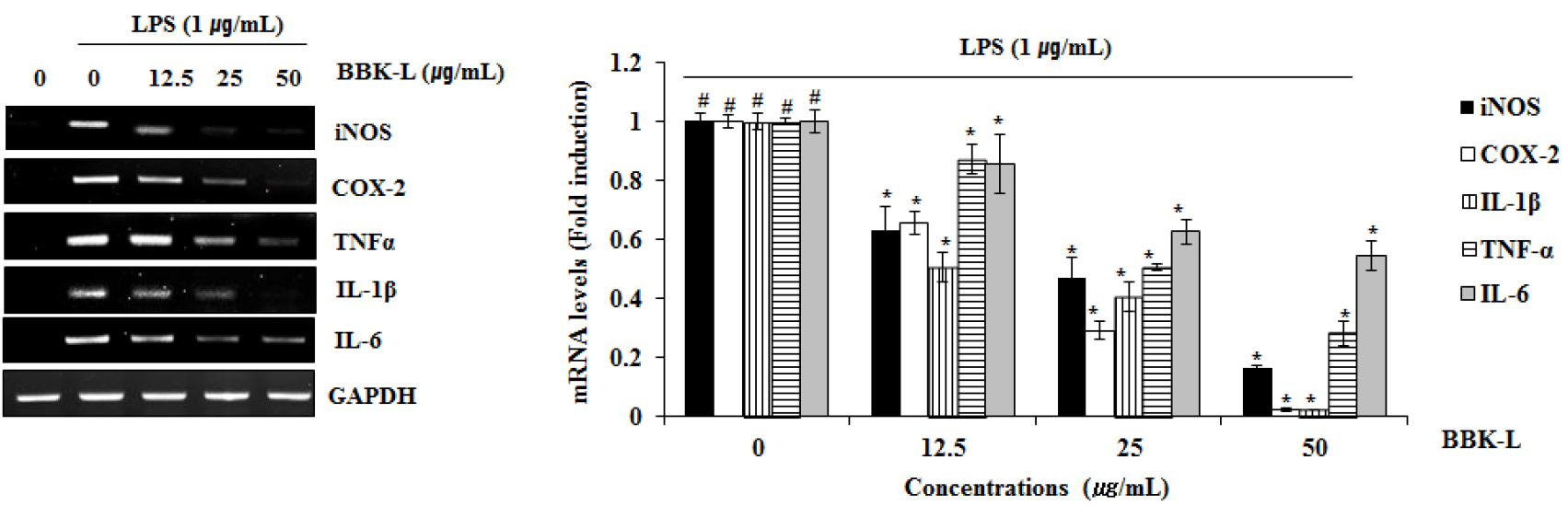
Fig. 2
Effect of BBK-L on the expression of iNOS, COX-2, TNF-α, IL-1β and IL-6 in LPS-stimulated RAW264.7 cells. RAW264.7 cells were pre-treated with BBK-L for 6 hours and then co-treated with LPS (1 μg/mL) for 18 hours. For RT-PCR analysis, total RNA was prepared. GAPDH was used as internal control for RT-PCR. #p < 0.05 compared to the cells without the treatment, and *p < 0.05 compared to the cells treated with LPS alone.
망개나무 잎 추출물의 NF-κB의 활성 억제 효과
BBK-L 추출물의 NF-κB 활성화 억제 효과를 평가한 결과, BBK-L 추출물을 농도 (12.5, 25, 50 ㎍/mL) 의존적으로 IκB-α의 분해 및 인산화를 억제하였다(Fig. 3A, B). 또한, NF-κB의 p65 핵 내 전이에 미치는 영향을 평가한 결과, BBK-L 추출물은 농도 의존적으로 핵 내 p65를 감소하는 경향을 보였다(Fig. 3C). LPS 자극에 의해 세포질 내에서 IκB가 인산화되면 IκB와 결합하고 있던 p65가 핵 안으로 이동하여 전사인자로써 작용하게 되므로 여러 염증성 사이토카인과 염증인자들의 발현을 유도하여 p65의 인산화가 세포질에서 핵으로의 이동을 조절하기 때문에 p65 인산화 저해는 NF-κB 활성 억제와 연관이 있는 것으로 알려져 있다(Hsieh et al., 2011; Kim et al., 2020; Moynagh, 2005). 따라서 BBK-L 추출물은 IκB-α의 인산화 및 분해를 억제하고, p65 핵 내 전이를 억제하여 항염증 효과를 나타내는 것으로 나타났다.
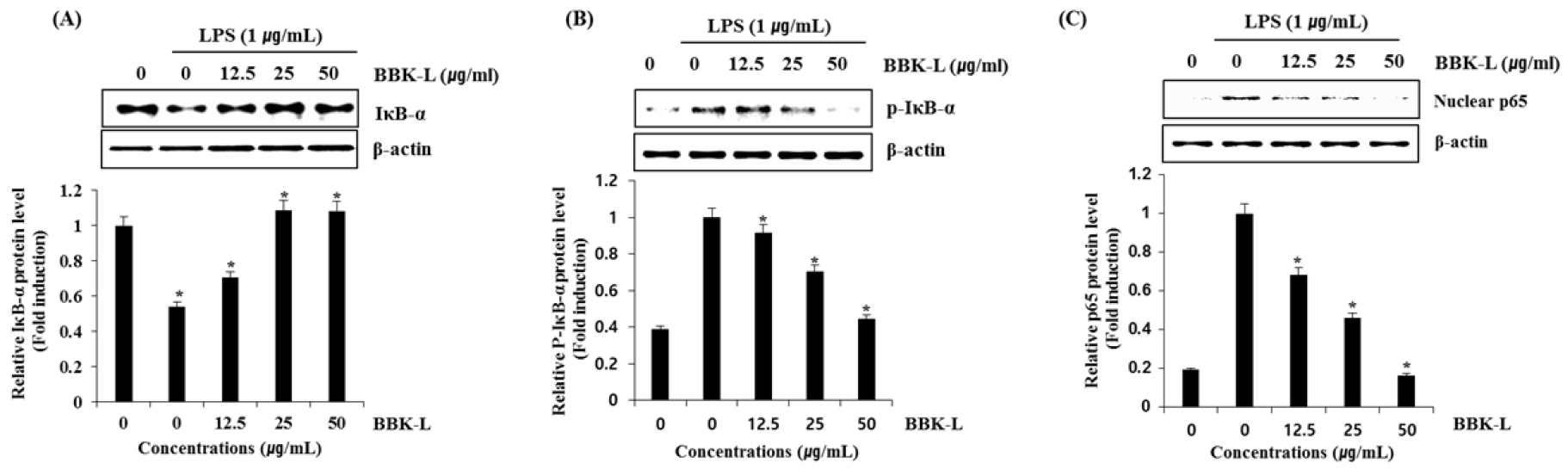
Fig. 3
Effects of BBK-L on NF-κB signaling activation in LPS-stimulated RAW264.7 cells (A) RAW264.7 cells were pretreated with BBK-L (0, 12.5, 25, 50 μ g/mL) for 6 hours and then co-treated with LPS (1 μg/mL) for 40 minutes. (A and B) RAW264.7 cells were pretreated for 6 hours with the indicated concentrations of BBK-L (12.5, 25, 50 μg/mL) and than co-treated LPS (1 μg/mL) treatment for 40 minutes. (C) RAW264.7 cells were pre-treated with BBK-L (0, 12.5, 25, 50 μg/mL) for 40 minutes. After the treatment, the cytosol and nucleus were prepared. For Western blot analysis, the cell lysates were subjected to SDS-PAGE and the Western blot was performed using antibodies against IκB-α and p-IκB-α and nucleus p65. β-actin was used as a internal control. #p < 0.05 compared to the cells without the treatment, and *p < 0.05 compared to the cells treated with LPS alone.
망개나무 잎 추출물의 MAPK의 활성 억제 효과
MAPK의 활성화는 각각 인산화를 통해 이루어지며 LPS에 의해 유도된 RAW264.7 세포에서 BBK-L 추출물로부터 MAPK의 인산화를 조절하는지 평가하기 위해서 농도별(0, 12.5, 25, 50 ㎍/mL)로 처리한 결과, p38은 인산화 억제 활성이 없는 것으로 나타났으나 ERK1/2, JNK의 인산화는 BBK-L 추출물에 의해서 억제되는 것을 확인하였다(Fig. 4). MAPK는 세포의 성장과 분화, cytokine과 스트레스에 대한 세포 반응의 조절에 중요한 역할을 한다고 알려져 있다(Ilorton et al., 1999). MAPK의 종류에는 p38, ERK1/2, JNK가 있으며 이들은 LPS에 의해 자극된 대식세포에서 염증 매개체의 합성과 분비를 조절하는데 관여하고 있다(Coskun et al., 2011; Zhang et al., 2014). 따라서 BBK -L 추출물은 ERK1/2, JNK의 인산화 조절을 통해 MAPK 활성을 저해함으로써 항염증 활성이 있다고 판단된다.
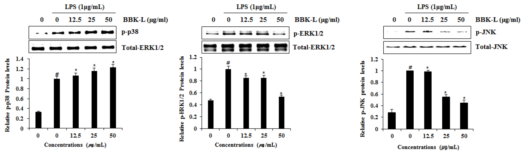
Fig. 4
Effects of BBK-L on MAPK signaling activation in LPS-stimulated RAW264.7 cells. RAW264.7 cells were pretreated with BBK-L (0, 12.5, 25, 50 μg/mL) for 6 hours and then co-treated with LPS (1 μg/mL) for 40 minutes. For western blot analysis, the cell lysates were subjected to SDS-PAGE and the western blot was performed using antibodies against p-p38, total-ERK1/2, p-ERK1/2, total p-38, p-JNK and total-JNK. #p < 0.05 compared to the cells without the treatment, and *p < 0.05 compared to the cells treated with LPS alone.
적 요
본 연구는 망개나무 추출물의 항염증 효과를 구명하기 위하여 수행되었다. 망개나무를 이용한 다양한 기능성 연구가 진행되고 있으나 망개나무 항염증에 대한 연구는 과학적 근거가 부족한 실정이다. 따라서 망개나무 잎(BBK-L) 추출물이 마우스 대식세포인 RAW264.7 세포에서 LPS에 의한 염증반응에 미치는 영향을 평가하였다. LPS로 유도된 마우스 대식세포 RAW264.7에 BBK-L 추출물을 농도별로 처리하였을 때, NO 생성량과 만성염증 유발인자인 iNOS, COX-2, IL-1β, TNF-α 및 IL-6 발현 감소를 확인하였으며, NF-κB 및 MAPK의 인산화를 억제하는 것을 확인하였다. 이러한 연구결과를 토대로 BBK-L 추출물이 만성 염증질환의 예방과 치료 또는 기능성 소재로 활용될 수 있는 기초적인 정보를 제공할 수 있을 것으로 사료된다.
