

Introduction
Materials and Methods
Preparation of BOE
Reagents and kits
Animal model
Histologic experiments
Statistical analysis:
Results and Discussion
Introduction
Major effective compound of B. oleracea, sulforaphane (4-methylsulfinylbutyl isothiocyanate) has been well identified as the most potent naturally occurring inducer of phase II enzymes (Fahey et al., 1997; Yoshida et al., 2015). Especially, dietary sulforaphane is known to ameliorators to various injuries such as carbon tetrachloride-induced intestinal ischemia reperfusion etc (Baek et al., 2008). In these reports, the ability of orally ingested sulforaphane-riched BOE to induce ameliorative effect was suggested as the basis for protection from these injuries.
Sulforaphane is a metabolite of glucoraphanin via hydrolyzation to sulforaphane by myrosinase (Bones and Rossiter, 1996; Zhang and Talalay, 1998). B. oleracea sprouts have been reported to induce detoxification enzymes in vitro and in vivo (Zhang et al., 2006). In other hands, B. oleracea synthesized naphthoquinone derivatives such as vitamin K families such as phylloquinone (Blanco and Blanco, 2017) by various plants including cabbage, cauliflower, tomatoes, spinach, and soybean, olive, and sunflower oils. Indeed, many phytochemicals were reported many medicinal effects such as cancer (Mokhtari et al., 2018; Ravichandiran et al., 2019), metabolic disorder (Teng et al., 2019), muscle inflammation (1), atherosclerosis (Shehatou and Suddek, 2016) and temporal/sustained neurodegeneration (Klomparens and Ding, 2019; Oh et al., 2013). Fortunately, few reports have evaluated already the effects of ingestion of B. oleracea sprouts on liver function via various mechanisms such as regulation of ER stress (Lei et al., 2018) and activation of nuclear erythroid 2-related factor 2 (Nrf2) (Xu et al., 2019), protection of inflammation in hepatic tumorigenesis (Chen et al., 2016). However, no has been reported on the improvement of lipidemic liver disorder with alcohol metabolism in the blood. Therefore, we investigate the effects of BOE on HFD and alcohol-induced liver with in vivo model. These reports show that alcoholic liver injuries are reduced by intake of BOE in in vitro and in vivo;
Materials and Methods
Preparation of BOE
To obtain BOE, B. oleracea were germinated from guaranteed seed and sprouts (3 days after budding) (KOREGON, Ansung, Korea) (20 ㎏) extracted with ten-fold volume of 70% ethanol at 40-60°C for 12 h. The extract was filtered by 0.45 ㎛ pore size filter (EMD Millipore, MA, USA) and concentrated with rotary evaporator (N-1300E, EYELA, Tokyo, Japan). Finally, the concentrated extract was sterilized by filtration through MF-Millipore filters (EMD Millipore, MA, USA), freeze dried (Bondero, IlShinBioBase Co Ltd, Korea), packed in plastic tubes (Nunc, CA, USA), and stored at -80°C (Thermo-Fisher, CA, USA) until use (Anydoctor HealthCare Co. Ltd. Seoul, Korea).
Reagents and kits
All guaranteed reagents were purchased from Sigma-Aldrich Co (MO, USA). Venous whole blood was taken from inferior vena cava, and then serum samples were sent to the Green Cross Laboratories (GC lab, GiHeung, Korea) on the same day. Parameters including hepatic enzymes such as alanine aminotransferase (ALT), alkaline phosphatase (ALP), and gamma-glutamyl transferase (GGT) as well as the lipid profile including, total cholesterol (T-CHO), triglyceride (TG), low-density lipoprotein cholesterol (LDL-C), and blood ethanol concentration were examined.
Animal model
We performed alcohol-induced fatty liver models (approval No WKU-17-67, Ethics Committee of the Wonkwang University). For the experiment using the ethanol-induced fatty liver model, male Sprague-Dawley rats aged 6 weeks were purchased from Orient Bio (Sungnam, Korea). After the acclimatization period, rats were divided into Normal control (n = 10), fatty liver model (n = 10), fatty liver model (high fat diet for 4 weeks) + 30 ㎎/㎏ BOE (n = 10), fatty liver model + 100 ㎎/㎏ BOE, and fatty liver model + 300 ㎎/㎏ BOE (n = 10) groups. Control and fatty liver groups were fed the normal diet, and the BOE treat groups were fed the BOE diet for 4 weeks. Fatty liver was induced in the fatty liver model (high fat diet + ethanol) group and fatty liver + BOE groups by oral administration of 6 mL/㎏ ethanol (15 mL/㎏ as 40% ethanol, v/v). The normal group was orally administered same solvent.
Histologic experiments
Histological experiments were conducted following common methods. Briefly, the dissected liver tissue was fixed in 10% formalin solution and embedded with paraffin. The paraffin sections (4-6 ㎛thickness) and stained with hematoxylin and eosin (H&E) following routine methods. Images were taken in randomly chosen microscopic views from individual slide of each animals using a microscope (TE-2000, Nikon, Tokyo, Japan)(Xie et al., 2018). Slides were graded for steatosis according to the Kleiner’s methods (Kleiner et al., 2005). A score of steatosis grade was assigned to describe the extent of lipid accumulated spaces in the hepatocytes (grade 0: <5%; grade 1: 5–33%; grade 2: 33–66%; grade 3: >66%) (Wang et al., 2015).
Statistical analysis:
All data are expressed as the Mean ± S.E.M. A one-way analysis of variance (ANOVA) test employed followed by Tukey’s multiple range tests to compare each group. Student’s t-test was used for comparisons of two groups. Statistical analyses conducted using SPSS for Windows software (Ver 10.0, Chicago, IL, USA) and data with different superscript letters are significantly different when p value is less than 0.05.
Results and Discussion
Single treatment of BOE to hepatocytes cell line had low cytotoxic effect on the concentration of dietary intake in rats (data not shown). The levels of body weight gain food intake, and water intake after HFD (control group) and HFD + EtOH administration (ethanol group), shown no significantly changed in the various dose of BOE group compared with each group (Fig. 1). Indeed, administration of different dose of BOE also shown no significant changes between each group. Whereas, the levels of ALP, ALT as a blood enzyme marker of functional integrity on liver were markedly reduced by administration of BOE in a dose dependent manner (Fig. 2). In other study, B. oleracea sprout extract-containing diet induced low increases of body weight than normal diets (Kikuchi et al., 2015; Lai et al., 2008). Similarly, we also show no significant changes of weight gain in the animals fed with direct administration of BOE (Fig. 1A). Therefore, these results suggested that BOE has ameliorative effects on HFD- and EtOH-induced liver disorder and BOE had effects to more different mechanisms to other organs, but may require long-term administration of BOE for observe of chronic effects, if necessary.
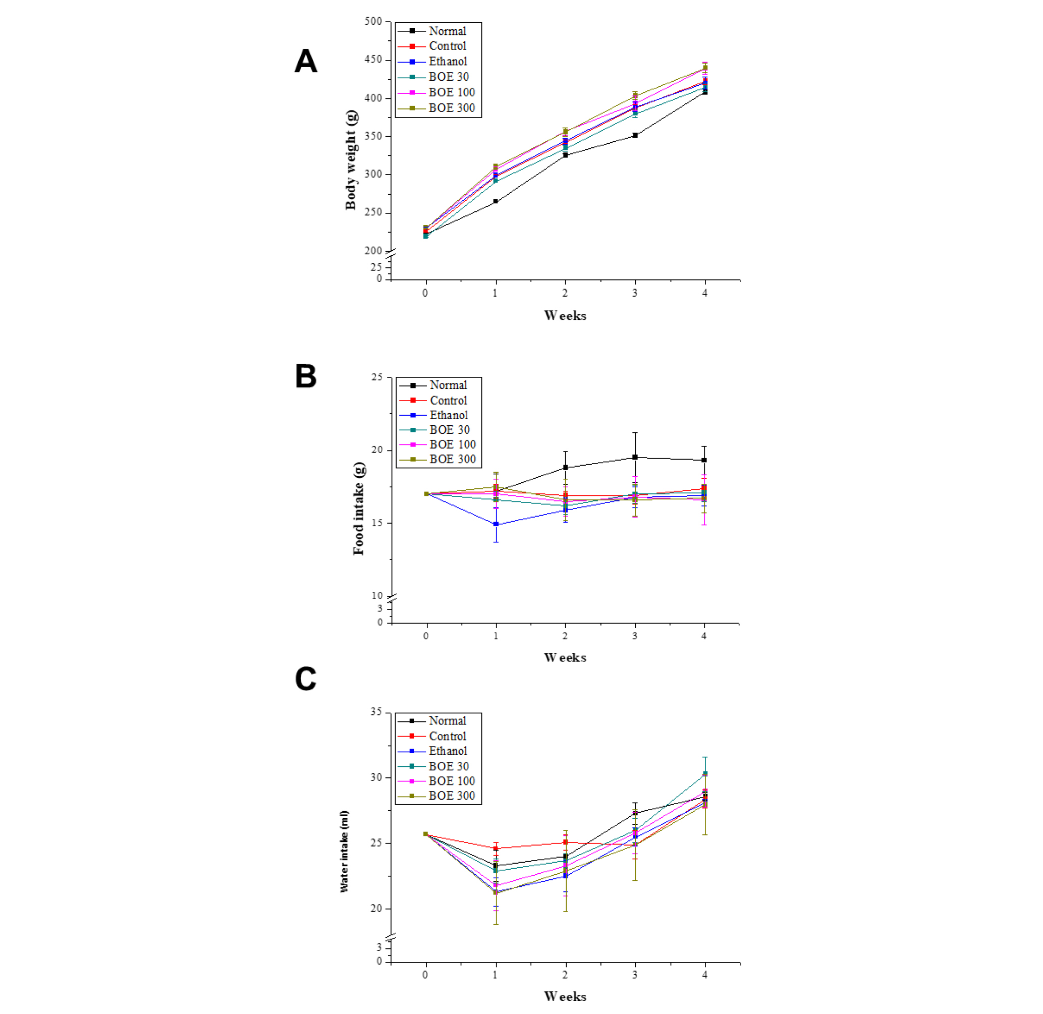
Fig. 1.
Changes of general profiles of BOE-administrated rats. After 4 weeks with normal diet (normal group) or high fat diet (control and ethanol group), BOEs were treated indicated dosages. (A) Body weight (B) food intake, and (C) water intake profiles were monitored and drawn as the line-scatter graphs. a,b,c Values in the row with different superscript letters are significantly different by Tukey’s multiple range tests at P < 0.05. Data are shown as mean ± SEM.
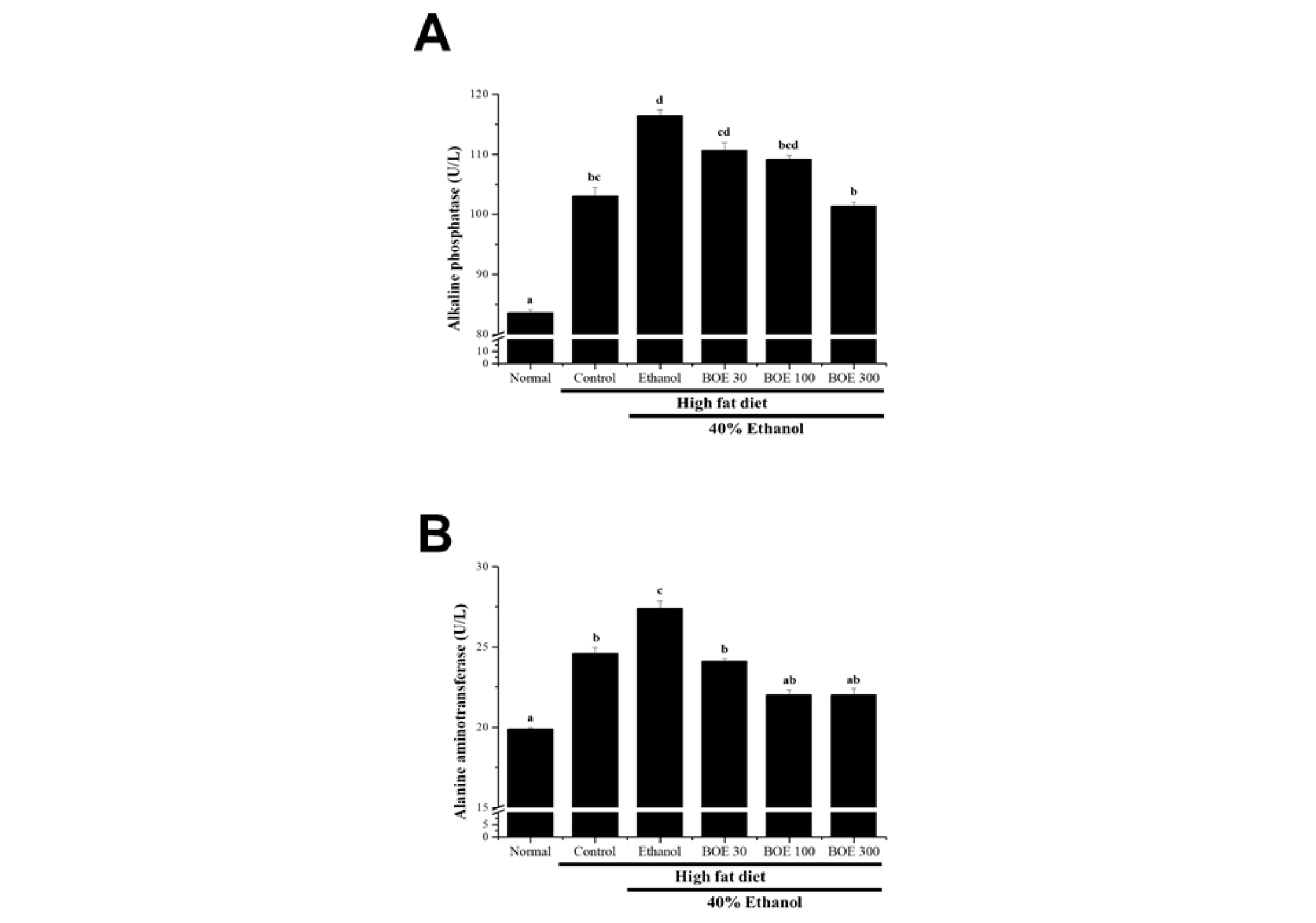
Fig. 2.
Changes of liver-related enzymes profiles in serum on HFD/alcohol-induced fatty liver model by BOE-administration. Normal diet (normal group) or high fat diet (control and ethanol group), BOEs were treated indicated dosages for 4 weeks and then measured (A) alkaline phosphatase (ALP) and (B) alanine aminotransferase (ALT). a,b,c Values in the row with different superscript letters are significantly different, P < 0.05. Data are shown as mean ± SEM.
Taken together, we measured blood levels of T-CHO, LDL-C, and TG (Fig. 3). In control and EtOH groups were significantly increased these lipid markers in blood, whereas administration of BOE was recovered compared with the control and/or EtOH group. Although, LDL-C was reduced by administration BOE in a dose dependent manner (Fig. 3B), however T-CHO and TG were shown lower dose dependency (Fig. 3A and C). Indeed, the level of high-density lipoprotein cholesterol (HDL-C) shows no significant changes in any groups (data now shown). Therefore, we need further examines specific mechanisms of BOE on lipid metabolism on same model in future.
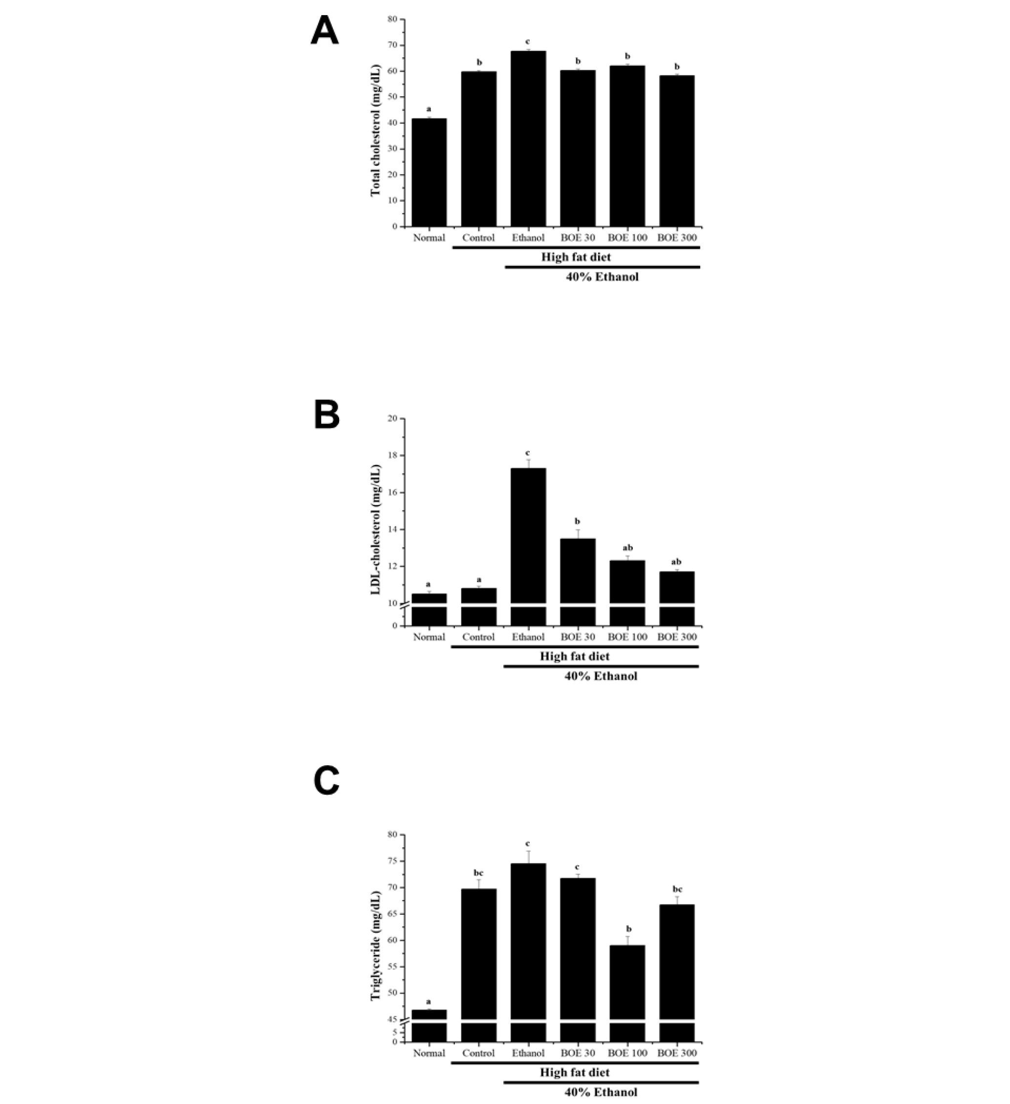
Fig. 3.
Changes of blood lipid profiles on HFD/alcohol- induced fatty liver model by BOE-administration. (A) Total cholesterol (T-CHO), (B) low-density lipoprotein cholesterol (LDL-C), and (C) triglyceride (TG) levels in serum. a,b,c Values in the row with different superscript letters are significantly different by Tukey’s multiple range tests at P < 0.05. Data are shown as mean ± SEM.
Taken together, we investigated effects of BOE administration on alcohol degradation in blood. Levels of blood alcohol were highly elevated at 1hr after EtOH administration. Interestingly, chronic administration of BOE were significantly reduced blood alcohol levels at same time point in a dose dependent manner and decreased area under curve (AUC) (Fig. 4). These results suggested that improving effects of BOE in animal liver function were also related against to alcohol metabolism.
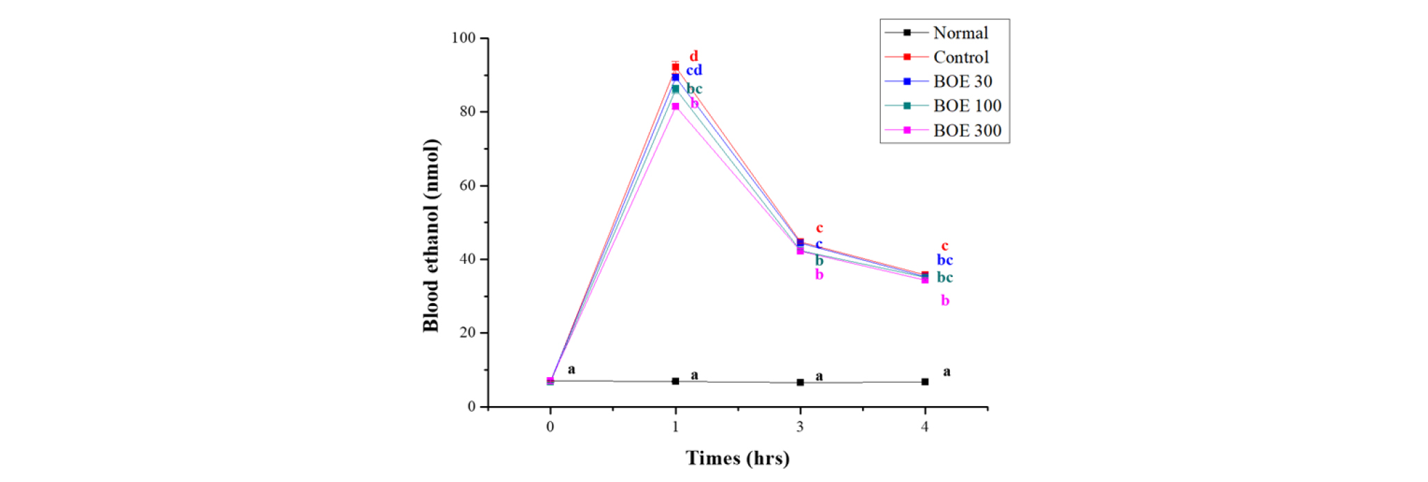
Fig. 4.
Effects of BOE on reduction of blood alcohol concentration on the HFD/alcohol-induced fatty liver. Ethanol was single administrated to both group and measured blood alcohol described in Materials and Methods. a,b,c Values in the row with different superscript letters are significantly different by Tukey’s multiple range tests at P < 0.05. Data are shown as mean ± SEM.
Based on these data, we performed histological experiments for evaluate that improving effects against HFD- and alcohol-induced fatty liver and its complication. Liver steatosis, abnormal retention of lipids within cells, is primary lipid metabolic problem. As a result, high fat diet and ethanol administration were markedly increased lipid and architecture malformation in liver tissue. In contrast, BOE administration were significantly reduced lipid droplet area in a dose dependent manner (Fig. 5) and relatively higher degree of steatosis accompanied by the presence of various size fat vacuoles (Fig 5B). These results suggest that administration BOE have ameliorative effects alcohol- and HFD-induced hepatic steatosis via reduction of lipid accumulation in hepatocytes.
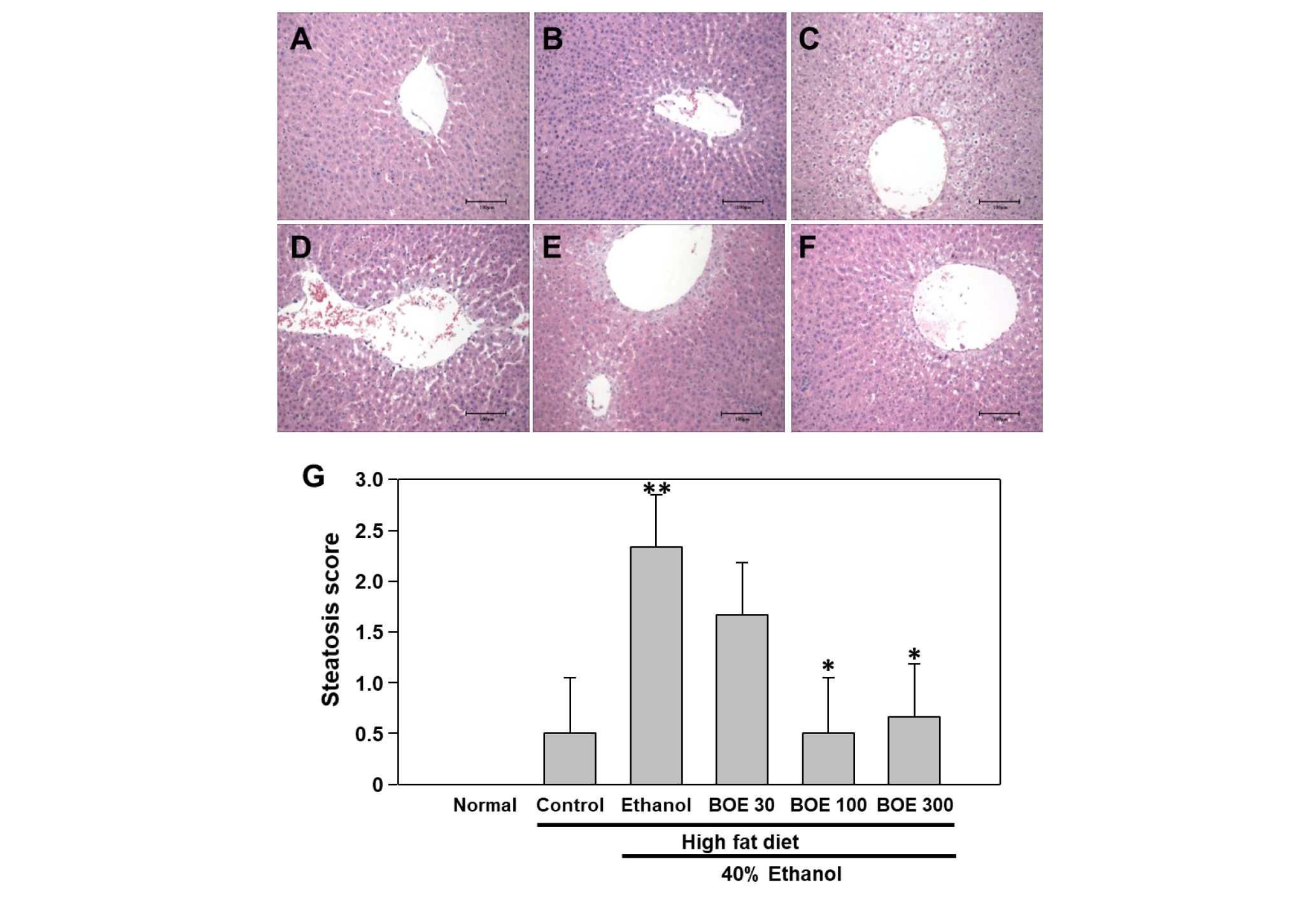
Fig. 5.
Ameliorative effects of BOE on high fat diet- and HFD/alcohol-induced fatty liver model. Tissue sections slide of (A) normal, (B) HFD, (C) HFD + EtOH, (D) HFD + EtOH + BOE 50 ㎎/㎏, (E) HFD + EtOH + BOE 100 ㎎/㎏, and (F) HFD + EtOH + BOE 300 ㎎/㎏ were stained with H<E methods and presented (G) steatosis scores. Scale bar = 100㎛. Magnification: ×20. ** indicates signficantly difference between normal and EtOH group by Student’s T-test at P < 0.01. * indicates significantly difference with HFD+EtOH group by Student’s T-test at P < 0.05. Data are shown as mean ± SEM.
In summary, we showed that BOE ameliorated liver functions and lipid metabolism in normal and fatty liver with EtOH administration in rat model. Moreover, BOE improved ability alcohol metabolism in rats model also. Our data suggest that protection from various liver injuries resulted from induction of anti-oxidation such as GSH synthesis and GST activity. Indeed, some other mechanisms may contribute to suppression of HFD and/or alcohol-induced liver injury with other liver problem (Xie et al., 2017). Therefore, we conclude that BOE had defensive and protected function to the hepatic-disorders of various types of excess lipid accumulation and alcohol consumption through acceleration of metabolism and antioxidation in the liver.
