

서 언
재료 및 방법
시료 추출
세포배양 및 세포생존률 측정
지방세포 분화유도 및 lipid accumulation 측정
Triglyceride contents
Western blot
통계처리
결과 및 고찰
세포생존률
세포내 lipid accumulation
세포내 triglyceride contents
세포내 중성지방 조절 유전자 발현
적 요
서 언
인체는 탄수화물, 지방 및 단백질 등의 영양소 섭취로 에너지 소비 및 저장를 통해 생존하지만 에너지의 공급이 소비보다 많아지면 과잉된 영양소는 지방으로 전환되어 지방세포에 저장되거나 간에서 중성지방으로 전환되어 소비된다(Bae et al., 2013; Bandyopadhyay et al., 2017). 간에서 지방은 지단백질의 형태로 혈액으로 배출되어 대사되지만 혈액 내 중성지방의 수치가 과도하게 높아지게 되면 고중성지방혈증 및 이상지질혈증의 원인이 되며 관상동맥질환과 같은 심혈관질환, 당뇨병, 고혈압 등 대사증후군의 원인이 되어 사회적 문제로 대두되고 있다(Kopelman, 2000; Park et al., 2015; Robert and Eckel, 2008).
이상지질혈증의 치료제는 콜레스테롤의 합성을 억제하는 statin과 PCSK9 inhibitor인 repatha (evolocumab), praluent (alirocumab) 등이 있으며, 콜레스테롤 흡수를 억제하는 ezetimibe, mRNA 기반 치료제인 inclisiran, LDL receptor의 특정 도메인을 모방한 small peptide 등이 있다(Bandyopadhyay et al., 2017; Kim et al., 2019). 그러나 사용되고 있는 치료제는 고가이거나 제2형 당뇨병, 인지기능 저하, 근육 약화와 같은 부작용이 발생되거나 상용화되지 못하고 있는 실정이다(Montan et al., 2019; Wicinski et al., 2017).
현대인들은 건강에 대한 관심이 고조되면서 식품섭취를 통해 질병 예방 또는 관리하고자 하는 소비자의 니즈가 커지면서 천연물을 이용한 건강기능식품 및 의약품의 사용이 증가하고 있다(Choung et al., 2013; Kim et al., 2012; Seo et al., 2013). 식품의약품안전처에 등재된 기능성 원료 중 혈중 중성지방 개선 소재로는 디글리세라이드 함유유지, 정제오징어유, 글로빈 가수분해물, EPA 및 DHA함유 유지, 난소화성말토덱스트린이 있다. 디글리세라이드 함유유지는 트리글리세라이드로 재합성되지 않아 혈중 중성지방 수치가 상승하지 않으며(Hong et al., 2009), 정제오징어유와 EPA 및 DHA함유 유지는 간에서 중성지방 합성을 감소시킴으로써 혈중 중성지방 수치를 감소시킨다(Choi, 2018). 난소화성 말토덱스트린은 식이섬유의 하나로 당과 지방의 소화 흡수를 억제시켜 혈중 중성지방을 감소시키며(Chung, 2019), 글로빈 가수분해물은 혈중 triglyceride와 chylomicron 감소, 소화관에서의 지방 흡수 감소, 간의 triglyceride lipase 활성 증가를 통해 중성지방을 분해시킨다고 보고되어 있다(Kagawa et al., 1995). 그러나 천연물을 이용한 혈중 중성지방 개선 연구는 아직 미흡한 실정으로 혈중 중성지방 개선 소재에 대한 추가 연구가 필요하다.
레몬밤(Melissa officinalis)은 지중해 연안에서 주로 자생하며 레몬향과 맛을 지닌 꿀풀과 식물로서 rosmarinic acid, geraniol, citronella, phenolic acid, terpene, caffeic acid 등의 유효성분이 함유되어 있다(Kennedy et al., 2006). 레몬밤은 항우울, 항산화, 항스트레스, 인지능 향상, 알츠하이머 완화, 면역 증진 등의 효과가 있다고 알려져 있으며(Emamghoreishi and Talebianpour, 2009; Ulbricht et al., 2004; Kennedy et al., 2003; Ramanauskiene et al., 2016), 식품의약품안전처에 등재된 건강기능식품 원료로 체지방감소에 도움을 줄 수 있는 개별인정형 소재로 인정되어 있다. 하지만 레몬밤 추출물의 혈중 중성지방 개선 효능에 대한 연구는 아직 미비한 실정이므로 본 연구에서는 레몬밤 추출물이 지방세포 내 lipid accumulation 및 TG 함량, ATGL, HSL, CGI-58, perilipin에 미치는 영향을 확인해 혈중 중성지방 개선 기능성 소재로의 가능성을 확인하고자 하였다.
재료 및 방법
시료 추출
실험에 이용된 레몬밤은 물 추출(MOW100), 70% 에탄올추출(MOE70), 50% 에탄올추출(MOE50), 30% 에탄올추출(MOE30) 조건으로 추출하였다. 레몬밤 추출은 무게 당 10배 추출용매를 각각 가한 뒤 60℃에서 5시간 shanking incubator를 이용하여 추출하였다. 5시간 후 실온에서 방냉한 뒤 3,000×g에서 30분간 원심분리하고 상층액을 Whatman filter paper (Whatman International, Maidstone, UK)로 여과한 후, 감압농축기(Eyela SB-1000, Tokyo, Japan)로 농축하여 동결건조기(FD 8508, Ilshin, Korea)로 건조하여 실험에 사용하였다.
세포배양 및 세포생존률 측정
3T3-L1 지방전구세포는 american type culture collection (ATCC, Manassas, USA)로부터 구입하여 10% bovine calf serum (Welgene, Daegu, Korea)과 1% penicillin-streptomycin (Welgene, Daegu, Korea)이 첨가된 dulbecco’s modified eagle’s medium (DMEM, Welgene, Daegu, Korea) 배지에서 37℃, 5% CO2 조건하에서 배양하였다. 레몬밤 추출물이 3T3-L1 세포에 미치는 독성평가는 3-[4,5-dimethylthiazole-2-yl]-2,5-di-phenyl- tetrazolium bromide (MTT)환원 방법을 이용하여 측정하였다. 분화되지 않은 3T3-L1 세포를 96 well plate에 1×10⁵ cells/mL로 100 μL씩 분주하고 24시간 배양한 뒤 FBS가 첨가되지 않은 배지에 소재를 희석하여 세포에 처리 후 24시간 배양하였다. 소재가 포함된 배양배지를 제거하고 MTT (0.5 ㎎/mL)를 배양배지에 녹여 100 μL씩 분주한 뒤 37℃에서 4시간 동안 배양한 후 배양배지를 제거하였다. 각 well에 형성된 formazan에 DMSO 100 μL를 첨가한 후 shaker를 이용하여 녹이고, 30분 후 UV/vis spectrophotometer (Opiwen 2120UV plus, Mecasys Co. Ltd., Korea)를 사용하여 570 ㎚에서 흡광도를 측정하였다. 대조군의 흡광도 값을 기준으로 세포독성을 확인하였다.
지방세포 분화유도 및 lipid accumulation 측정
3T3-L1 지방전구세포를 지방세포로 분화하기위해 6 well plate에 5×10⁵ cell/well의 세포를 분주하여 세포가 완전히 밀집되게 배양하였고 2일을 더 배양한 후 MDI (0.5 mM 3-isobutyl-1-methylxanthine (IBMX), 0.5 uM dexamethasone, 10 ㎍/mL insulin) solution과 10% FBS을 포함하는 DMEM 배지에서 2일 동안 배양함으로써 분화를 개시한 다음, 10 ㎍/ml insulin 및 10% FBS를 포함하는 DMEM 배지로 교환하여 2일 동안 분화를 진행시켰다. 그 이후로는 10% FBS만을 포함하는 DMEM 배지에서 4일 동안 배양함으로써 세포 내 지방축적에 의해 지방구(lipid droplet)를 형성하는 지방세포로 분화를 완료하였다. Lipid accumulation 측정은 MDI solution 및 배지교환시마다 레몬밤 추출물을 0.05, 0.1, 0.2 ㎎/mL 농도로 동시에 처리해 분화유도가 완료되는 시점까지 배양하였다. 분화유도가 완료된 세포는 PBS로 2회 세척하고 10% formalin을 처리하고 4℃에서 1시간 동안 고정시킨 뒤 세척하고 60% isopropanol solution을 처리해 지방세포를 염색하였다. 염색된 세포는 PBS로 세척하고 100% isopropanol을 이용해 oil red O를 용출한 뒤 520 ㎚에서 흡광도를 측정해 대조군과 비교해 lipid accumulation을 확인하였다.
Triglyceride contents
Triglyceride contents 측정은 EZ-Triglyceride Quantification Assay Kit를 이용해 분석하였다. 3T3-L1 지방전구세포를 지방세포로 분화하기 위해 6 well plate에 5×10⁵ cell/well의 세포를 분주하여 세포가 완전히 밀집되게 배양하였고 2일을 더 배양한 후 MDI (0.5 mM 3-isobutyl-1-methylxanthine (IBMX), 0.5 uM dexamethasone, 10 ㎍/mL insulin) solution과 10% FBS을 포함하는 DMEM 배지에서 2일 동안 배양함으로써 분화를 개시한 다음, 10 ㎍/mL insulin 및 10% FBS를 포함하는 DMEM 배지로 교환하여 2일 동안 분화를 진행시켰다. 그 이후로는 10% FBS만을 포함하는 DMEM 배지에서 4일 동안 배양함으로써 세포 내 지방축적에 의해 지방구(lipid droplet)를 형성하는 지방세포로 분화를 완료하였다. Lipid accumulation 측정은 MDI solution 및 배지교환시마다 레몬밤 추출물을 0.05, 0.1, 0.2 ㎎/mL 농도로 동시에 처리해 분화유도가 완료되는 시점까지 배양하였다. 분화유도가 완료된 세포는 PBS로 2회 세척하고 NP40을 이용해 균질시킨 뒤 100℃에서 가열해 세포내 존재하는 triglycerol을 용해시키고 13,000 rpm에서 2분간 원심분리해 불용성 물질을 제거하였다. Lysate 및 standard는 96 well plate에 50 μL와 lipase 2 μL를 넣어주고 실온에서 20분간 반응시킨 뒤 triglyceride assay buffer 46 μL, triglyceride enzyme mix 2 μL, triglyceride probe 2 μL를 각각 넣어준 뒤 실온에서 30분간 반응시킨 뒤 570 ㎚에서 흡광도를 측정해 대조군과 비교해 TG contents를 확인하였다.
Western blot
분화가 완료된 3T3-L1 지방세포에 레몬밤 추출물을 0.2 ㎎/mL 농도로 처리하고 24시간 배양하였다. 배양이 완료된 세포는 cell scraper로 긁어 모으고 , 5,000 rpm에서 10분간 운심분리하고 RIPA buffer를 이용해 lysis하였다. Lysis된 세포는 bradford 방법으로 정량한 동일양의 단백질을 sodium dodecyl sulfate (SDS)와 β-mercapto-ethanol를 포함한 sample buffer에 3:1로 혼합한 후 100℃에서 10분간 가열하였다. 단백질 샘플은 SDS-PAGE로 전기영동한 후 polyvinylidene fluoride membrane (0.45 μm, PVDF transfer membrane, Thermo, Rockford, IL, USA)으로 transfer하였다. Membrane은 0.1% tween 20과 5% skim milk를 함유한 tris-buffered saline (TBS)에 2시간 동안 blocking 하였다. 그 후 PKA (1:1000), phospho-PKA (1:1000), p-hormone sensitive lipase(Ser563) (1:1000), HSL (1:1000), β-actin (1:2000)(Cell signaling technology, danvers, MA, USA), HSL (Ser563)(1:1000)(Thermo Fisher, MA, USA) 1차 antibody가 첨가된 buffer에서 overnight동안 반응하고 TBS-T (TBS containing 0.1% tween 20)로 5분씩 3회 세척하였다. 그런 다음 horseradish peroxidase-conjugated secondary antibody (1:1000)(Santa Cruz Biotechnology, Santa Cruz, CA, USA)가 첨가된 buffer에서 1시간 동안 반응한 후 enhanced chemiluminescence method를 이용하여 x-ray 필름에 감광시켰다. Detection된 밴드의 강도는 imageJ (National institutes of health, Bethesda, MD, USA) 소프트웨어를 이용하여 분석하였다.
통계처리
통계처리는 SAS (statistical analysis system) program을 사용하여 각각의 시료에 대한 평균±표준편차로 나타내었으며, 각 군의 평균치의 통계적 유의성을 p < 0.05 수준에서 ANOVA test 및 Duncan's multiple range test로 사후분석 후 유의성을 검정하였다.
결과 및 고찰
세포생존률
레몬밤 추출물이 3T3-L1 preadipocyte에 미치는 독성을 확인하기 위해 MOW100, MOE70, MOE50, MOE30을 농도별로 3T3-L1 세포에 처리하고 24시간 후 MTT assay를 통해 확인한 결과는 Fig. 1과 같다. 레몬밤 추출물을 3T3-L1 preadipocyte에 0.01, 0.25, 0.5, 1, 2, 5 ㎎/mL 농도로 24시간 배양하였을 때 MOW100은 5 ㎎/mL 농도까지 처리하여도 세포독성을 나타내지 않았으나 MOE70, MOE50, MOE30는 2, 5 ㎎/mL 농도로 처리하였을 때 80% 이하의 세포생존률을 나타내어 preadipocyte에 독성을 나타내는 것이 확인되었다.
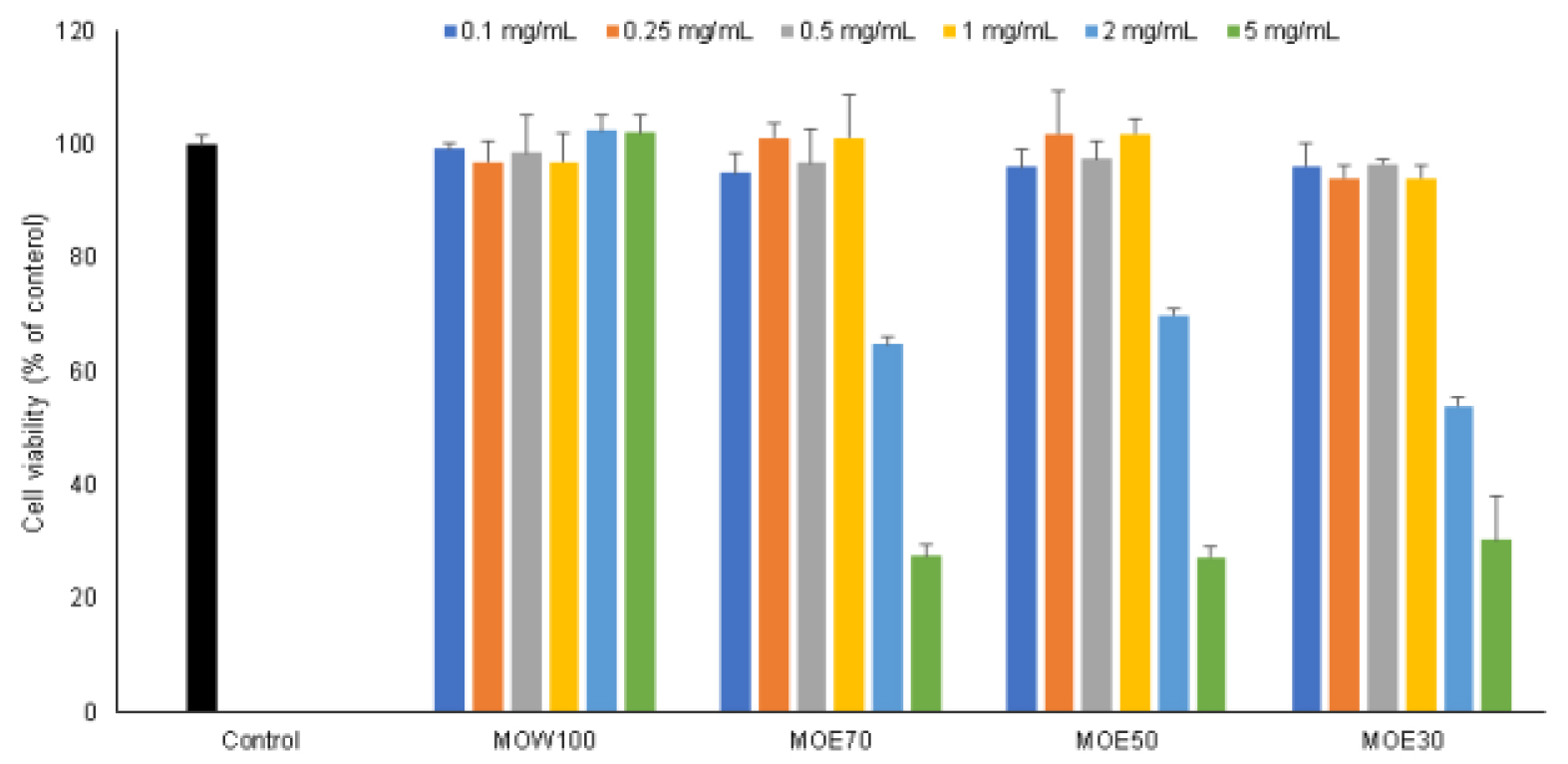
Fig. 1.
Concentration-dependent effects of Melissa officinalis extract on 3T3-L1 cell viability. Cell viability was analyzed using MTT assay. Values expressed are mean±SD of triplicate measurements. Values with different superscripts in column are significantly different (P < 0.05) by Duncan's multiple range test.
세포내 lipid accumulation
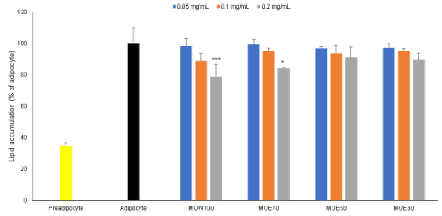
3T3-L1 지방전구세포는 지방세포로 분화가 가능한 세포주로 지방세포 분화과정에서 형태의 관찰과 더불어 지방합성 및 분해를 조절하는 유전자발현에 대한 연구가 가능하다(Baek et al., 2013; Feng et al., 2014). 레몬밤 추출물이 지방세포 분화유도과정에서 lipid accumulation에 미치는 영향을 확인한 결과는 Fig. 2와 같다. 실험결과 MOW100의 경우 0.05 ㎎/mL 처리시 1.6%, 0.1 ㎎/mL 처리시 11.0%, 0.2 ㎎/mL 처리시 21.3%의 lipid accumulation을 억제시키는 것이 확인되었으며, MOE70의 경우 0.05 ㎎/mL 처리시 0.6%, 0.1 ㎎/mL 처리시 4.8%, 0.2 ㎎/mL 처리시 15.8%의 lipid accumulation를 억제시키는 것이 확인되었다. MOE50의 경우 0.05 ㎎/mL 처리시 3.1%, 0.1 ㎎/mL 처리시 6.3%, 0.2 ㎎/mL 처리시 8.9%의 lipid accumulation을 억제시키는 것이 확인되었으며, MOE30의 경우 0.05 ㎎/mL 처리시 2.8%, 0.1 ㎎/mL 처리시 4.8%, 0.2 ㎎/mL 처리시 10.6%의 lipid accumulation를 억제시키는 것이 확인되었다. 레몬밤 추출물 중 물 추출 조건인 MOW100는 에탄올 추출조건인 MOE70, MOE50, MOE30보다 세포내 lipid accumulation를 억제 효능이 높은 것으로 확인되었다.
Lee et al. (2020)와 Woo et al. (2016)의 연구결과에서 레몬밤 추출물 처리시 3T3-L1 지방세포내 lipid accumulation 억제시켰으며, 추출조건에 따른 레몬밤 추출물의 효능 검증을 위해 세포내 TG 및 western blot 실험으로 레몬밤 추출물의 중성지방 조절 효능을 검증하였다.
세포내 triglyceride contents
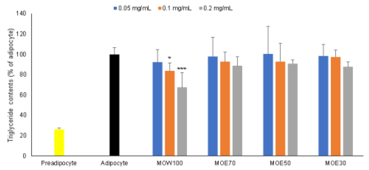
지방세포는 분화시 세포내 tiglyceride를 축적시키며, 글리세롤과 지방산으로 구성된 TG는 비만 및 고지혈증의 원인이 된다(Listenberger et al., 2003). 레몬밤 추출물이 지방세포 분화유도과정에서 TG에 미치는 영향을 확인한 결과는 Fig. 3와 같다. 실험결과 MOW100의 경우 0.05 ㎎/mL 처리시 7.7%, 0.1 ㎎/mL 처리시 16.5%, 0.2 ㎎/mL 처리시 32.7%의 TG 축적을 억제시키는 것이 확인되었으며, MOE70의 경우 0.05 ㎎/mL 처리시 2.0%, 0.1 ㎎/mL 처리시 7.4%, 0.2 ㎎/mL 처리시 11.5%의 TG 축적을 억제시키는 것이 확인되었다. MOE50의 경우 0.1 ㎎/mL 처리시 7.1%, 0.2 ㎎/mL 처리시 9.2%의 TG 축적을 억제시키는 것이 확인되었으며, MOE30의 경우 0.05 ㎎/mL 처리시 1.5%, 0.1 ㎎/mL 처리시 2.7%, 0.2 ㎎/mL 처리시 12.4%의 TG 축적을 억제시키는 것이 확인되었다. 레몬밤 추출물 중 물 추출 조건인 MOW100는 에탄올 추출조건인 MOE70, MOE50, MOE30보다 세포내 TG 축적 억제 효능이 높은 것으로 확인되었다.
고지혈증 환자가 레몬밤 분말을 섭취하였을 때 LDL-cholesterol과 triglyceride가 감소된다고 보고하였으며(Parisa et al., 2016), 이러한 결과는 3T3-L1 지방전구세포가 지방분화 과정에 레몬밤 추출물이 triglyceride 축적 억제 실험결과와 일치함이 확인되었다.
세포내 중성지방 조절 유전자 발현
3T3-L1 지방전구세포는 지방세포로 분화되면서 지방 생성 및 대사를 통해 중성지방을 합성하며 분화가 진행될수록 중성지방은 lipid droplet의 형태로 결합되며(Aboulaich et al., 2011; Yu et al., 2021), 이렇게 형성된 중성지방은 각종 호르몬 및 교감신경의 작용에 의해 유리지방산(fatty acid)과 글리세롤(glycerol)로 가수분해 된다(Frayn et al., 2003). 중성지방이 분해과정의 하나로 adrenergic receptor-adenylate cyclase에 의해 조절되는 cAMP 경로가 주요한 중성지방 분해기전으로 알려져 있으며, PKA, HSL, perilipin, ATGL 및 CGI-58에 의해 중성지방의 분해가 이루어진다(Brasaemle, 2007; Holm, 2003; Londos et al., 1995).
PKA는 cAMP 의존적으로 조절되며 세포내 cAMP 증가에 의해 p-Thr172 위치가 인산화되면서 HSL 및 perilipin 인산화를 통해 lipolysis를 조절한다(Holm, 2003; Rayalam et al., 2008). Fig. 4의 (b)는 PKA 단백질 발현 실험결과로 MOW100 처리시 152.9%, MOE70 처리시 136.7%, MOE50 처리시 126.7%, MOE30 처리시 124.7% 증가시키는 것이 확인되었다.
HSL는 PKA, AMPK 등에 조절받는 lipase중 하나로 인슐린(insulin)이나 에피네프린(epinephrine) 등의 호르몬에 의해 활성화된 PKA에 의해 인산화된다. HSL은 p-Ser563과 p-Ser660을 비롯한 다양한 인산화 위치가 존재하는데 p-Ser563 인산화는 HSL의 직접적인 활성화를 유도하고 p-Ser660 인산화를 지방구로의 전위를 유도한다. Perilipin은 lipid drople에서 발견되며 지방분해를 방해하는 작용을 하며, HSL과 상호 작용을 통해 유리지방산(free fatty acid) 및 글리세롤(glycerol)의 분해가 조절한다(Carmen and Victor, 2006; Holm, 2003; Rayalam et al., 2008). Fig. 4의 (c)와 (d)는 HSL 단백질 발현 실험결과로 p-Ser563 HSL 단백질 발현은 MOW100 처리시 153.0 %, MOE70 처리시 106.4%, MOE50 처리시 73.6%, MOE30 처리시 92.6%로 나타났으며, p-Ser660 HSL 단백질 발현이 103.1%, MOE70 처리시 94.7%, MOE50 처리시 89.3%, MOE30 처리시 85.6%로 나타났다. Fig. 4의 (e)는 perilipin 단백질 발현 실험결과로 MOW100 처리시 88.0 %, MOE70 처리시 96.6%, MOE50 처리시 98.7%, MOE30 처리시 106.3%로 나타났다.
ATGL은 지방세포에서 주로 발현되는 lipase 중 하나로 triglyceride를 diglyceride로 직접적으로 분해하는 속도제한 지방분해효소로 보고된 바 있으며(Haemmerle et al., 2006; Zechner et al., 2005), CGI-58은 perilipin과 복합체를 이루고 있다가 perilipin이 인산화되면 분리가 되는데 떨어져나온 CGI-58은 ATGL과 복합체를 형성해 ATGL의 활성화를 유도한다(Greenberg et al., 2011). Fig. 4의 (f)는 ATGL, (g)는 CGI-58 단백질 발현 실험결과로 MOW100 처리시 102.2 %, MOE70 처리시 116.7%, MOE50 처리시 104.5%, MOE30 처리시 119.8%로 나타났으며, CGI-58 단백질 발현은 MOW100 처리시 122.1%, MOE70 처리시 121.6%, MOE50 처리시 90.84%, MOE30 처리시 93.1%로 나타났다.
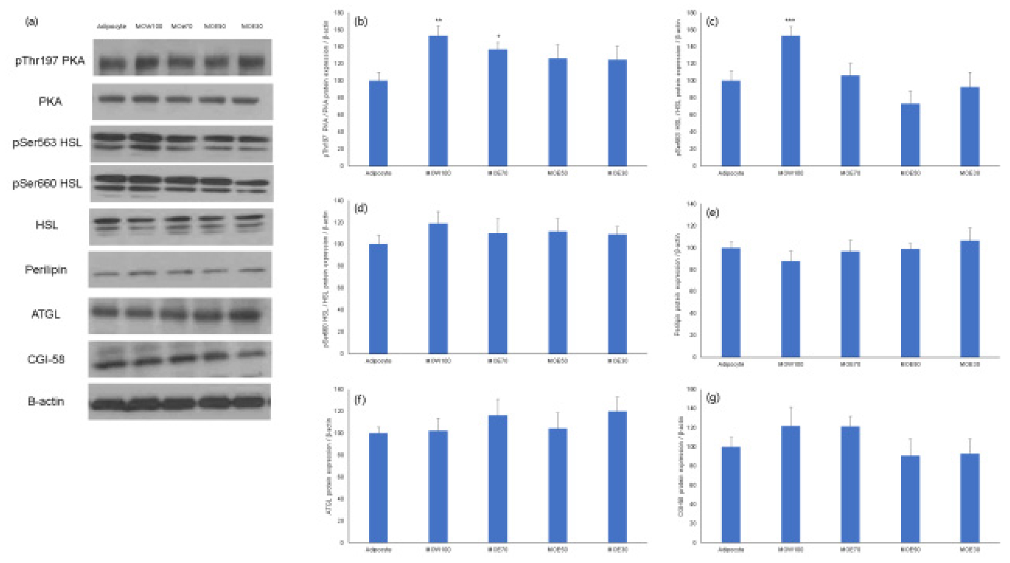
Fig. 4.
Effect of Melissa officinalis extract on lipolysis-related protein expression. (a) western blot band, (b)pThr197 PKA/PKA, (c) pSer563 HSL/HSL, (d) pSer660 HSL/HSL, (e) perilipin, (f) ATGL, (g) CGI-58 analyzed by Image J. Values expressed are mean±SD of triplicate measurements. Values with different superscripts in column are significantly different (P < 0.05) by Duncan's multiple range test.
적 요
본 연구는 추출용매에 따른 레몬밤 추출물의 중성지방 조절에 대한 논문으로 3T3-L1 지방전구세포를 이용해 지방세포로 분화유도한 뒤 lipid accumulation, triglyceride contents, PKA, HSL, perilipin, ATGL 및 CGI-58 단백질 발현을 확인하였다. 실험결과 네가지 조건의 레몬밤 추출물 중 물 추출 조건인 MOW100 추출물만 lipid accumulation과 triglyceride contents의 유의적 억제 효능을 나타내었으며, MOE70, MOE50, MOE50 추출물은 lipid accumulation과 triglyceride contents의 감소효과가 없거나 유의적 차이를 나타내지 않는 것이 확인되었다. MOW100 추출물의 세포내 지방 축적 억제는 지방세포 내 lipolysis를 조절하는 효소 조절을 통해 나타난 결과로 판단된다. 따라서 레몬밤 물 추출물은 cAMP-PKA를 활성화시키고, lipolysis를 조절하는 효소의 상승작용을 통해 세포내 중성지방을 조절함으로써 혈중 중성지방을 개선하는 후보소재로의 사용이 가능할 것으로 사료된다.
